. 2009 Jan;20(1):379-88.
doi: 10.1091/mbc.e07-10-1007. Epub 2008 Nov 12.

Katanin knockdown supports a role for microtubule severing in release of basal bodies before mitosis in Chlamydomonas

Affiliations

Katanin knockdown supports a role for microtubule severing in release of basal bodies before mitosis in Chlamydomonas

M Qasim Rasi et al. Mol Biol Cell. 2009 Jan.

Abstract

Katanin is a microtubule-severing protein that participates in the regulation of cell cycle progression and in ciliary disassembly, but its precise role is not known for either activity. Our data suggest that in Chlamydomonas, katanin severs doublet microtubules at the proximal end of the flagellar transition zone, allowing disengagement of the basal body from the flagellum before mitosis. Using an RNA interference approach we have discovered that severe knockdown of the p60 subunit of katanin, KAT1, is achieved only in cells that also carry secondary mutations that disrupt ciliogenesis. Importantly, we observed that cells in the process of cell cycle-induced flagellar resorption sever the flagella from the basal bodies before resorption is complete, and we find that this process is defective in KAT1 knockdown cells.


Figures

Figure 1.
Figure 1.
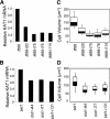
Isolation of two KAT1 knockdown strains. (A) pEZ-KAT1-RNAi construct used to generate stable RNAi lines. Cells transformed with this construct were sequentially selected on plates containing paromomycin, and then on plates containing Zeocin, to confirm the presence of the PsaD-driven hairpin targeting KAT1 (large inward-pointing arrows). (B) Box plots of cell volume distributions of two KAT1-RNAi isolates and the parental cw2 strain. Boxes encompass 50% of all data points (the interquartile range) and error bars encompass 90% of all data points. The boundary between the black and white boxes is the median cell volume. (C) Box plots of cell volumes of both KAT1-RNAi isolates after backcross to a wild-type (cell-walled) strain, as well as the wild-type cell volume. Boxes encompass the interquartile range and error bars encompass 90% of all data points. (D) Anti-acetylated tubulin immunofluorescence visualization of typical cells from each isolate (right) shows lack of flagella in both KAT1-RNAi isolates. Bars, 7.5 μm (cw2) and2.5 μm (KAT1-RNAi#1 and KAT1-RNAi#4). (E) Quantification of mRNA from cw2 and the two KAT1-RNAi strains. Main graph shows relative KAT1 level (the amount of KAT1 mRNA in cw2 cells is set at 1). The inset graph shows relative control transcript (RPS14) level. (F) Agarose gel electrophoresis of reverse transcriptase-PCR products. Amplicons targeted putative genes disrupted by insertion of KAT1-RNAi constructs into strain KAT1-RNAi#4 (left) and KAT1-RNAi#1 (right). All cDNA pools showed accumulation of PCR products targeting a control gene (data not shown).
Figure 2.
Figure 2.
KAT1 can be readily knocked down in flagella-less cells. (A) Relative KAT1 mRNA level, as determined by real-time PCR, from a representative subset of ift88 strains transformed with, and selected for, pEZ-KAT1-RNAi. (B) Relative KAT1 mRNA level, as determined by real-time PCR, from a representative subset of bld1 strains transformed with, and selected for, pEZ-KAT1-RNAi. (C) Box plots of cell volume distribution of the same ift88-KAT1-RNAi isolates examined in A. Error bars capture 90% of cells. (D) Box plots of cell volume distribution of the same bld1-KAT1-RNAi isolates examined in A. Error bars capture 90% of cells.
Figure 3.
Figure 3.
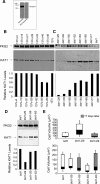
KAT1 protein can be readily knocked down in bld1 mutants. (A) Characterization of anti-KAT1 antiserum. Wild-type cell lysate was blotted in duplicate and probed with either immune antiserum (left) or preimmune serum from the same rabbit (right). The expected molecular weight of KAT1 is 60 kDa. (B) Quantification of relative knockdown of KAT1 protein levels by anti-KAT1 Western blot (middle) in seven random isolates recovered from transformation of wild-type cells with pEZ-KAT1-RNAi. Anti-PKG2 was used as a loading control by reprobing the same blot (top). Gel films were scanned and densitometric analysis (bottom) performed as described in Materials and Methods. (C) Quantification of relative knockdown of KAT1 protein levels by anti-KAT1 Western blot (middle) in seven random isolates recovered from transformation of bld1 cells with pEZ-KAT1-RNAi. Anti-PKG2 was used as a loading control by reprobing the same blot (top). Gel films were scanned and densitometric analysis (bottom) performed as described in Materials and Methods. (D) Quantification of relative knockdown of KAT1 protein levels in a subset of the same isolates examined in C after an additional 2 wk of growth. Analysis as described for C. (E) Box plots showing cell volumes of the same bld1:KAT1-RNAi isolates examined in B and C. White boxes show cell volume distribution data from the time of the experiment shown in B and gray boxes are data from the time of the experiment shown in C. Boxes encompass the interquartile range and error bars encompass 90% of the total data points.
Figure 4.
Figure 4.
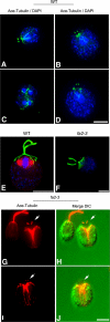
Basal bodies are freed from flagella by severing before mitosis. (A–D) Representative images of flagellar remnants associated with representative wild-type mitotic cells. Remnants are visible as dots staining for acetylated-α-tubulin (green) and are found at the anterior end of the cell (A, B, and D) or in a position consistent with the prior anterior of the mother cell, in cells that have completed at least one round of division (C). Blue is 4,6-diamidino-2-phenylindole (DAPI), which stains both nuclear and plastid DNA. (E) Longer flagella (stained with anti-acetylated tubulin; green) are observed more rarely in wild-type mitotic cells. The spindle of this cell visualized with anti-α-tubulin (red). (F–J) Remnants are found in mitotic fa2 mutant cells. (F) a fa2 cell early in cell division has relatively long flagella as stained by acetylated tubulin (green), counterstained with DAPI (blue). (G and I) fa2 cells later on in cell division, as indicated by the presence of cleavage furrows, have flagellar remnants as visualized by acetylated-tubulin staining (red; the primary antibody for these cells was a mixture of anti-acetylated- and anti-α-tubulin). (H and J) Overlay with corresponding DIC images. Bars, 5 μm.
Figure 5.
Figure 5.
DIC images of presumptive cw2:KAT1-RNAi cells. (A–C) Examples of presumptive KAT1 knockdown cells in a cw2, cell wall-less background. Note the presence of flagella on dividing cells. Similar cells were not seen in any control colonies. Bar, 5 μm.
Figure 6.
Figure 6.
Severing may occur at either end of the flagellar transition zone. Deflagellation in response to stress involves severing at the distal end of the transition zone (the SOFA), after which the flagellum is cast away from the cell, whereas the transition zone remains associated with the basal body and can nucleate the assembly of a new flagellum. Resorption before mitosis involves both shortening of the flagellum from its distal end and severing at the proximal transition zone. This proximal severing frees the basal body, leaving the transition zone. Any residual flagella, or flagellar remnants, remain associated with the cell wall. AX, axoneme; TZ, transition zone; BB, basal body.

Similar articles

Cited by

References

    1. Bradley B. A., Quarmby L. M. A NIMA-related kinase, Cnk2p, regulates both flagellar length and cell size in Chlamydomonas. J. Cell Sci. 2005;118:3317–3326. - PubMed
    1. Brazelton W. J., Amundsen C. D., Silflow C. D., Lefebvre P. A. The bld1 mutation identifies the Chlamydomonas osm-6 homolog as a gene required for flagellar assembly. Curr. Biol. 2001;11:1591–1594. - PubMed
    1. Brodersen P., Sakvarelidze-Achard L., Bruun-Rasmussen M., Dunoyer P., Yamamoto Y. Y., Sieburth L., Voinnet O. Widespread translational inhibition by plant miRNAs and siRNAs. Science. 2008;320:1185–1190. - PubMed
    1. Buster D., McNally K., McNally F. J. Katanin inhibition prevents the redistribution of gamma-tubulin at mitosis. J. Cell Sci. 2002;115:1083–1092. - PubMed
    1. Cavalier-Smith T. Basal body and flagellar development during the vegetative cell cycle and the sexual cycle of Chlamydomonas reinhardii. J. Cell Sci. 1974;16:529–556. - PubMed

Publication types

LinkOut - more resources