. 2009 Jan 15;457(7227):313-7.
doi: 10.1038/nature07487. Epub 2008 Nov 12.

Experience leaves a lasting structural trace in cortical circuits

Affiliations

Experience leaves a lasting structural trace in cortical circuits

Sonja B Hofer et al. Nature. .

Abstract

Sensory experiences exert a powerful influence on the function and future performance of neuronal circuits in the mammalian neocortex. Restructuring of synaptic connections is believed to be one mechanism by which cortical circuits store information about the sensory world. Excitatory synaptic structures, such as dendritic spines, are dynamic entities that remain sensitive to alteration of sensory input throughout life. It remains unclear, however, whether structural changes at the level of dendritic spines can outlast the original experience and thereby provide a morphological basis for long-term information storage. Here we follow spine dynamics on apical dendrites of pyramidal neurons in functionally defined regions of adult mouse visual cortex during plasticity of eye-specific responses induced by repeated closure of one eye (monocular deprivation). The first monocular deprivation episode doubled the rate of spine formation, thereby increasing spine density. This effect was specific to layer-5 cells located in binocular cortex, where most neurons increase their responsiveness to the non-deprived eye. Restoring binocular vision returned spine dynamics to baseline levels, but absolute spine density remained elevated and many monocular deprivation-induced spines persisted during this period of functional recovery. However, spine addition did not increase again when the same eye was closed for a second time. This absence of structural plasticity stands out against the robust changes of eye-specific responses that occur even faster after repeated deprivation. Thus, spines added during the first monocular deprivation experience may provide a structural basis for subsequent functional shifts. These results provide a strong link between functional plasticity and specific synaptic rearrangements, revealing a mechanism of how prior experiences could be stored in cortical circuits.


Figures
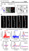
Figure 1
Figure 1. Monocular deprivation in adult mice increases the rate of spine addition in layer 5 neurons in binocular cortex
a, Timeline of the experimental protocol. b, Schematic of the mouse visual system (left), intrinsic signal map of the binocular visual cortex (middle, scale bar: 500 μm) and low-magnification image of a L5 neuron apical dendrite (right, scale bar: 50 μm). c, OD shifts during contralateral eye MD, 1-2 weeks after eye re-opening (BV: binocular vision) and during a second MD, measured by intrinsic signal imaging and shown as ratio of contralateral to ipsilateral eye response strength (contra/ipsi ratio). Circles depict data from individual mice, horizontal lines indicate mean values. d, High-magnification view of the dendritic stretch shown in b (red box), imaged every 4 days (depth ~25 μm, soma depth ~625 μm). MD in the contralateral eye was induced at the end of the third imaging session (day 8). Arrows point to spines appearing (red) or disappearing (blue) compared to the previous imaging session. Scale bar: 5 μm. e-h, Percentage of spines appearing (spine gain) and disappearing (spine loss) on layer 5 neurons between two imaging time points plotted against time for different cortical regions or conditions: binocular visual cortex (e, left, middle: individual neurons; right: average data, 22 cells, 13 mice, 2360 spines), monocular region of primary visual cortex (f, 7 cells, 6 mice, 754 spines), border of the binocular region (g, 9 cells, 9 mice, 984 spines), and control data from non-deprived mice (h, 21 cells, 11 mice, 2468 spines). Error bars denote SEM.
Figure 2
Figure 2. Monocular deprivation in adult mice does not alter spine dynamics in layer 2/3 neurons
a, Repeatedly imaged dendritic stretch of a layer 2/3 pyramidal neuron (imaging depth ~50 μm, soma depth ~210 μm) shown in b (side view) and c (top view, red box outlines dendritic stretch shown in a, scale bar: 100 μm). Arrows mark spine changes from previous imaging time point (red: spine gained, blue: spine lost). Scale bar: 5 μm. d, Average spine gain and loss on layer 2/3 pyramidal neurons in binocular visual cortex (7 cells, 4 mice, 1101 spines). Error bars denote SEM.
Figure 3
Figure 3. MD-induced increase in spine density on layer 5 neurons outlasts the altered experience
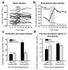
a, Spine density as a function of time for all imaged layer 5 neurons in binocular visual cortex (22 cells, 13 mice, 2360 spines). b, Average normalized spine density of layer 5 cells showing an increase in spine density during MD, imaged for at least 32 days (black, 14 cells, 9 mice, 1316 spines). c, Density of newly appeared spines that remain stable for a minimum of 16 days. The data have been split according to the time when the spines first appeared: before MD, during MD, and during equivalent periods in control animals. d, Ratio of persistent new spines to all new spines before and during MD. Matched control data from non-deprived mice are shown in gray. Error bars indicate SEM.
Figure 4
Figure 4. A second MD increases the size of spines gained during the first MD without additional spine gain
Rate of spine gain and loss (a,b) and normalized spine density (c,d) on layer 5 pyramidal neurons during a second period of contralateral-eye MD induced 2-3 weeks after the first (a,c, 14 cells, 9 mice, 1316 spines), and during a single MD in control mice of similar implant duration and number of imaging sessions before the MD (b,d, 14 cells, 11 mice, 1693 spines). e, Example of a spine that appeared during the first 4 days of MD and its brightness/size changes over time. BV: binocular vision. f, Average integrated brightness of persistent new spines that appeared during MD (red) or during the corresponding time period in non-deprived control animals (gray), normalized to the spine brightness value at the end of the first MD or the equivalent time point in control mice. Error bars indicate SEM.

Similar articles

Cited by

References

    1. Wiesel TN, Hubel DH. Single cell responses in striate cortex of kittens deprived of vision in one eye. J. Neurophysiol. 1963;26:1003–1017. - PubMed
    1. Clark SA, Allard T, Jenkins WM, Merzenich MM. Receptive fields in the body-surface map in adult cortex defined by temporally correlated inputs. Nature. 1988;332:444–445. - PubMed
    1. Hofer SB, Mrsic-Flogel TD, Bonhoeffer T, Hübener M. Prior experience enhances plasticity in adult visual cortex. Nat. Neurosci. 2006;9:127–132. - PubMed
    1. Bailey CH, Kandel ER. Structural changes accompanying memory storage. Annu. Rev. Physiol. 1993;55:397–426. - PubMed
    1. Yuste R, Bonhoeffer T. Morphological changes in dendritic spines associated with long-term synaptic plasticity. Annu. Rev. Neurosci. 2001;24:1071–1089. - PubMed

Publication types