. 2009 Mar;10(2):162-73.
doi: 10.1038/gene.2008.91. Epub 2008 Nov 13.

Dramatically reduced surface expression of NK cell receptor KIR2DS3 is attributed to multiple residues throughout the molecule

Affiliations

Dramatically reduced surface expression of NK cell receptor KIR2DS3 is attributed to multiple residues throughout the molecule

C J VandenBussche et al. Genes Immun. 2009 Mar.

Abstract

Using flow cytometry, fluorescent microscopy and examination of receptor glycosylation status, we demonstrate that an entire killer cell immunoglobulin-like receptor (KIR) locus (KIR2DS3)--assumed earlier to be surface expressed--appears to have little appreciable surface expression in transfected cells. This phenotype was noted for receptors encoded by three allelic variants including the common KIR2DS3*001 allele. Comparing the surface expression of KIR2DS3 with that of the better-studied KIR2DS1 molecule in two different cell lines, mutational analysis identified multiple polymorphic amino-acid residues that significantly alter the proportion of molecules present on the cell surface. A simultaneous substitution of five residues localized to the leader peptide (residues -18 and -7), second domain (residues 123 and 150) and transmembrane region (residue 234) was required to restore KIR2DS3 to the expression level of KIR2DS1. Corresponding simultaneous substitutions of KIR2DS1 to the KIR2DS3 residues resulted in a dramatically decreased surface expression. Molecular modeling was used to predict how these substitutions contribute to this phenotype. Alterations in receptor surface expression are likely to affect the balance of immune cell signaling impacting the characteristics of the response to pathogens or malignancy.


Figures

Figure 1
Figure 1. KIR2DS3 is minimally detected on the surface of NKL and Jurkat cells
KIR2DS1 and KIR2DS3 were expressed in NKL and Jurkat cells. a, Transfectants expressing KIR2DS1 (allele *002) and KIR2DS3 (allele *001) carrying N-terminal FLAG tags were surface-stained using anti-FLAG antibody and GFP-positive cells were analyzed by flow cytometry. Transfectants without FLAG-tags served as negative controls. NKL (b) and Jurkat (c) cells were transfected with KIR constructs encoding a C-terminal V5-tag in addition to the N-terminal FLAG-tag. Cells were surface-stained using an anti-FLAG antibody and intracellularly-stained using an anti-V5 antibody. Relative surface expression was normalized to FLAG-KIR2DS1 (by student’s t-test vs. control: *, P < 0.05, **, P<0.01; ***, P < 0.001; vs. FLAG-KIR2DS1: ^^^, P < 0.001; FLAG-KIR2DS3*001 and FLAG-KIR2DS3*002 vs. FLAG-KIR2DS3*004: #, P<0.05). In (b), levels of expression of KIR2DS3 receptors encoded by three allelic variants are compared to KIR2DS1 NKL cells transfected with V5-tagged-KIR2DS1 without a FLAG tag were a negative control. d, C-terminally V5-tagged KIR2DS1, KIR2DS3, or KIR2DL3 were expressed in NKL cells. After the biotinylation of surface proteins, receptors in cell lysates were immunoprecipitated using the anti-V5 antibody and analyzed by gel electrophoresis and Western blotting using anti-V5 (for total protein) or streptavidin (for surface biotinylated protein) probes. e, NKL cells were transfected with C-terminally V5-tagged KIR2DS1 or KIR2DS3. After staining the plasma membrane with DiO (green stain) and tagged protein with anti-V5 antibody (red stain), the images were merged to determine possible colocalization (shown as yellow).
Figure 2
Figure 2. Comparison of protein sequences of KIR2DS1 and KIR2DS3 identifies amino acids that may impact surface expression
Alignment of some of the allelic products of KIR2DS1 and KIR2DS3. The consensus and allelic KIR sequences were obtained from the IPD-KIR database . An asterisk at the 3′ end of the of the KIR2DS3*002 and KIR2DS3*004 allelic products indicates that the sequence is incomplete.
Figure 3
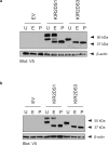
Figure 3. Digestion of KIR2DS1 and KIR2DS3 identifies a mature receptor isotype for KIR2DS1 but not KIR2DS3
NKL (a) or Jurkat (b) cells were transfected with empty vector (EV) or V5-tagged KIR2DS1 or KIR2DS3 and the cell lysates were mock digested (U) or digested with endoglycosidase H (E) or PNGase F (P). Receptors were analyzed by gel electrophoresis and Western blotting using the V5-specific antibody as a probe. β-actin monitors the amount of protein loaded on the gel.
Figure 4
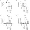
Figure 4. Two polymorphisms in the leader peptide significantly decrease KIR surface expression
Individual mutations were made at polymorphic positions in the leader peptide of the N-terminally FLAG-tagged, C-terminally V5-tagged KIR2DS1 receptor. The T(–18)M mutant converts the amino acid sequence from that encoded by KIR2DS1*002 to that encoded by a different allele at that locus, KIR2DS1*003. NKL (a) and Jurkat (b) cells were transfected with the mutated receptor genes and surface expression was analyzed by flow cytometry (vs. KIR2DS1 by student’s t-test: *, P < 0.05; ***, P < 0.001). NKL (c) and Jurkat (d) cells were transfected with N-terminally FLAG-tagged, C-terminally V5-tagged KIR2DS3 or KIR2DS3 mutant constructs for flow cytometric analysis of relative surface expression (vs. KIR2DS3 by student’s t-test: ^^, P < 0.01; ^^^, P < 0.001). In all instances (a-d), C-terminally V5-tagged KIR2DS1 without a FLAG tag was used as a negative control. Note that KIR2DS3 mutant expression levels in Figure 4 and subsequent figures are compared to wild type KIR2DS3 expression; these levels of expression are dramatically reduced compared to KIR2DS1. The level of KIR2DS1 expression in relationship to KIR2DS3 is shown in Figures 1b, 1c.
Figure 5
Figure 5. The D2 domain N123S and L150F polymorphisms significantly decrease KIR surface expression
NKL (a) and Jurkat (b) cells were transfected with N-terminally FLAG-tagged, C-terminally V5-tagged KIR2DS1, KIR2DS3, or KIR2DS1 mutant constructs for flow cytometric analysis of relative surface expression. The statistically significant loss-of-function mutations 123(N>S) and 150(L>F) were analyzed individually and in combination (vs. KIR2DS1 by student’s t-test: **, P < 0.01; ***, P < 0.001; vs. N123+L150F: ^^, P < 0.01; ^^^, P < 0.001). NKL (c,d) and Jurkat (e) cells were transfected with N-terminally FLAG-tagged, C-terminally V5-tagged KIR2DS3 or similarly tagged KIR2DS3 mutant constructs for flow cytometric analysis of surface expression (vs. KIR2DS3 by student’s t-test: *, P < 0.05; **, P < 0.01; ***, P < 0.001). Panels c and d show the same data. In panel c, the level of expression of the mutants is compared to KIR2DS3; in panel d, the level of expression is compared to KIR2DS1. The level of KIR2DS1 expression in relationship to KIR2DS3 is shown also in Figures 1b, 1c. For all flow cytometry studies, V5-tagged KIR2DS1 without a FLAG tag was used as a negative control. (f), NKL cells were transfected with empty (EV), V5-tagged KIR2DS1, or V5-tagged KIR2DS1 123(N>S) mutant expression vectors. Receptors from transfectant lysates were immunoprecipitated with an anti-V5 antibody and analyzed by gel electrophoresis and Western blotting using the same anti-V5 antibody as a probe . KIR2DS1 (g) and KIR2DS3 (h) were modeled based on the X-ray crystal structure of KIR2DS2. The residues at positions 123 and 150, which are critical for KIR surface expression, are shown.
Figure 6
Figure 6. The I234L polymorphism in the transmembrane domain decreases KIR surface expression in NKL cells
Individual mutations at polymorphic positions were made KIR2DS1 altering the transmembrane and intracellular domains of the N-terminally FLAG-tagged, C-terminally V5-tagged receptor. Relative surface expression of KIR in NKL (a) and Jurkat (b) cells was analyzed by flow cytometry (vs. KIR2DS1 by student’s t-test: *, P < 0.05; **, P < 0.01; ***, P < 0.001). Transfection of NKL (c) and Jurkat (d) cells with N-terminally FLAG-tagged, C-terminally V5-tagged KIR2DS3 or KIR2DS3 mutant constructs for flow cytometric analysis of surface expression (vs. KIR2DS3 by student’s t-test: **, P < 0.01). The level of KIR2DS1 surface expression in relationship to KIR2DS3 is shown in Figures 1b, 1c. In all instances (a-d), V5-tagged KIR2DS1 without an N-terminal FLAG tag was used as a negative control.
Figure 7
Figure 7. KIR2DS3 surface expression is greatly increased by five mutations made in combination
NKL (a) and Jurkat (b) cells were transfected with N-terminally FLAG-tagged, C-terminally V5-tagged KIR2DS1, KIR2DS3 or KIR2DS1 mutant constructs and relative surface expression was analyzed (vs. KIR2DS1 by student’s t-test: *, P < 0.05; **, P < 0.01; ***, P < 0.001; vs. KIR2DS3: ^, P < 0.05, ^^^, P < 0.001). NKL (c) and Jurkat (d) cells were transfected with N-terminally FLAG-tagged, C-terminally V5-tagged KIR2DS1, KIR2DS3, or KIR2DS3 mutant constructs for flow cytometric analysis of relative surface expression (vs. KIR2DS3 by student’s t-test: **, P < 0.01; ***, P < 0.001; vs. KIR2DS1: ^^, P < 0.01; ^^^, P < 0.001). In all instances (a-d), V5-tagged KIR2DS1 without a FLAG tag was used as a negative control.

Similar articles

Cited by

References

    1. Lanier LL. NK cell recognition. Annu Rev Immunol. 2005;23:225–274. - PubMed
    1. Hsu KC, Chida S, Dupont B, Geraghty DE. The killer cell immunoglobulin-like receptor (KIR) genomic region: gene-order, haplotypes and allelic polymorphism. Immunological Reviews. 2002;190(1):40–52. - PubMed
    1. Rajagopalan S, Long EO. Understanding how combinations of HLA and KIR genes influence disease. J Exp Med. 2005;201(7):1025–1029. - PMC - PubMed
    1. Stewart CA, Laugier-Anfossi F, Vely F, Saulquin X, Riedmuller J, Tisserant A, et al. Recognition of peptide-MHC class I complexes by activating killer immunoglobulin-like receptors. Proc Natl Acad Sci U S A. 2005;102(37):13224–13229. - PMC - PubMed
    1. Katz G, Markel G, Mizrahi S, Arnon TI, Mandelboim O. Recognition of HLA-Cw4 but not HLA-Cw6 by the NK cell receptor killer cell Ig-like receptor two-domain short tail number 4. J Immunol. 2001;166:7260–7267. - PubMed

Publication types