

Review
doi: 10.1038/nrc2523.
Epub 2008 Nov 13.
GammaH2AX and cancer
Affiliations
- PMID: 19005492
- PMCID: PMC3094856
- DOI: 10.1038/nrc2523
Item in Clipboard
Review
GammaH2AX and cancer
Nat Rev Cancer.
2008 Dec.
Abstract
Histone H2AX phosphorylation on a serine four residues from the carboxyl terminus (producing gammaH2AX) is a sensitive marker for DNA double-strand breaks (DSBs). DSBs may lead to cancer but, paradoxically, are also used to kill cancer cells. Using gammaH2AX detection to determine the extent of DSB induction may help to detect precancerous cells, to stage cancers, to monitor the effectiveness of cancer therapies and to develop novel anticancer drugs.
Figures
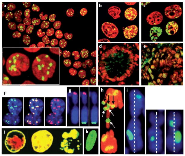
a | Human peripheral blood mononuclear cells 30 minutes after exposure to 1 Gy of ionizing radiation (IR). b | Colon cancer cells treated with campthotecin. c | Leukaemic cells treated with etoposide. d | Frozen sections of normal human colon. e | Frozen sections of colon adenocarcinoma. f | γH2AX foci (left) and p53 binding protein 1 (right) co-localize (middle) in mouse fibroblasts 30 minutes after exposure to 1 Gy of IR. g | Eroded telomeres in ageing cells. Telomeric DNA (left, red), γH2AX (right, green) and merged (middle). The putative eroded telomere (bottom) exhibits a γH2AX focus but is too short to bind the telomeric DNA probe, whereas the putative functional telomere (top) binds telomeric DNA and lacks a γH2AX focus. h | Broken metaphase chromosomes of Indian muntjac cells 90 minutes after exposure to 0.6 Gy of IR. The white arrows denote γH2AX foci on broken chromatid ends and the black arrows denote foci on the remaining metaphase chromosomes. i | Selected chromosomes of a WI38 normal human fibroblast metaphase spread 30 minutes after exposure to 1 Gy of IR. The left panel shows the largest chromosome in the metaphase, assumed to be chromosome 1 or chromosome 2, with a length about 245 Mbp (dotted white bar). The foci are approximately 40 and 16 Mbp. The middle and right panels show two other chromosomes from the same metaphase at the same scale. The chromosome in the middle panel has an estimated length of 169 Mbp and a focus about 20 Mbp. The chromosome in the right panel has an estimated length of 149 Mbp with foci of 45 and 22 Mbp. j | Colon cancer cells treated with TRAIL (TNF-related apoptosis-inducing ligand). From left to right: peripheral nuclear staining (1 hour treatment), pan-staining and apoptotic bodies fully stained with γH2AX (3 hour treatment). k | A normal human fibroblast after being exposed to an acute dose of ultraviolet C, exhibiting a pan-nuclear staining pattern. DNA is red for images a–e, h and j and blue for images f, g and i. γH2AX is green for all images. Part h is reproduced, with permission, from REF. © The Rockefeller University Press (1999).
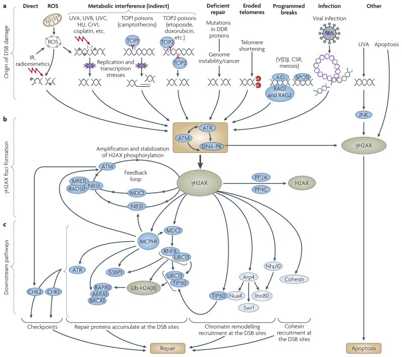
a | There are several categories of origins of DSBs. Direct: H2AX phosphorylation occurs after treatment with agents including ionizing radiation (IR) or radiomimetic drugs,. ROS are ions or small molecules (produced by normal metabolism and exogenous agents) that can cause DSBs. Indirect: Drugs, chemicals and DNA modifications induce replication and transcription stress, leading to DSBs,,. Ultraviolet (UV) radiation can cause DSBs in S-phase ,. Deficient repair: Mutations in DNA-damage repair (DDR) proteins result in genomic instability, leading to DSBs. Eroded telomeres: Critically short telomeres reveal double-strand ends,. Programmed: H2AX phosphorylation occurs in V(D)J recombination, class switch recombination (CSR) and meiosis,,. Infection: Retroviral integration induces DSBs. b | γH2AX focus growth. Three kinases, ataxia telangiectasia mutated (ATM), ataxia telangiectasia and Rad3-related (ATR) and DNA-dependent protein kinase (DNA-PK), respond to DSBs, resulting in initial H2AX phosphorylation,,. A signal amplification loop involving H2AX, nibrin (also known as NBS1) and mediator of DNA damage checkpoint protein 1 (MDC1) stimulates ATM and increases H2AX phosphorylation. Protein phosphatases PP2A and PP4C bind to and dephosphorylate γH2AX,, c | Downstream signalling pathways. NBS1 (REFS 139,140) and MDC1 (REF. 141) bind to γH2AX, which allows the accumulation of DDR proteins including the MRN (MRE11–RAD50–NBS1) complex, RNF8, BRCA1 and p53-binding protein 1 (53BP1),,. RNF8 catalyses the ubiquitylation of H2AX, which then recruits RAP80 (also| known as UIMC1) and BRCA1 to DSBs through ABRA1 (REF. 18). Accumulation at DSB sites of MCPH1 (also known as BRIT1) also depends on γH2AX. MCPH1 is involved in chromatin condensation and cell-cycle arrest, by interacting with BRCA1, NBS1, CHK1, 53BP1, MDC1, ATR, RPA (replication protein A) and ATM,. Accumulation of 53BP1, BRCA1, CHK1 and NBS1 on accrued γH2AX through MDC1 and/or MCPH1 could explain the role of γH2AX in checkpoints. Chromatin remodelling complexes participate in DNA repair. The human histone acetyltransferase TIP60 complex (in yeast, Nua4 complex) and the yeast ino80 and Swr1 complexes interact with H2AX or γH2AX,,. DSBs facilitate the association between TIP60 and UBC13 (also known as UBE2N), regulating the H2AX acetylation that is necessary for H2AX ubiquitylation (Ub) and its release from chromatin. γH2AX is crucial for sister-chromatid homologous recombination, probably by facilitating the interaction between sister chromatids,. H2AX has multiple roles during apoptosis,,,. Human proteins are represented in dark blue, yeast proteins in light blue. AID, activation-induced cytidine deaminase; CrVI, hexavalent chromium; HU, hydroxyurea; RAG, V(D)J recombination-activating protein; ROS, reactive oxygen species; TOP, topoisomerase.
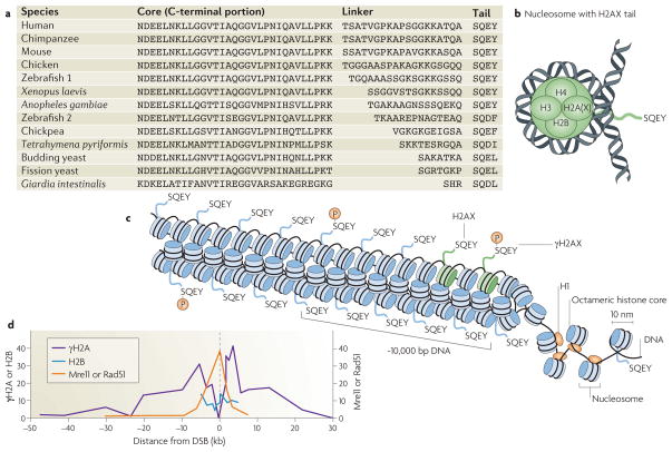
a | H2AX is an H2A histone with a core sequence conserved with other H2A species and a tail conserved through evolution connected by a linker of variable length. b | The SQEY tail extends from the core nucleosome near the entry and exit point of the DNA. c | The nucleosomes form a 30 nm fibre with H2AX molecules in every fifth nucleosome on average in mammals and every nucleosome in yeast. Approximately 10% of the H2AX molecules are phosphorylated at any one time in a focus. d | In yeast, chromatin immunoprecipitation studies show that H2A, the functional analogue of H2AX, is not phosphorylated near the site of the double-strand break (DSB), although histones (H2B) are present. Two repair proteins, Mre11 and Rad51, accumulate very near the break site. Part d modified, with permission, from REF. (2004) © Elsevier Ltd.
References
-
- McKinnon PJ, Caldecott KW. DNA strand break repair and human genetic disease. Annu Rev Genomics Hum Genet. 2007;8:37–55. - PubMed
-
- Jeggo PA, Lobrich M. DNA double-strand breaks: their cellular and clinical impact? Oncogene. 2007;26:7717–7719. - PubMed
-
- O’Driscoll M, Gennery AR, Seidel J, Concannon P, Jeggo PA. An overview of three new disorders associated with genetic instability: LIG4 syndrome, RS-SCID and ATR–Seckel syndrome. DNA Repair (Amst) 2004;3:1227–1235. - PubMed
Publication types
MeSH terms
Substances
Grants and funding
LinkOut - more resources
Full Text Sources
Other Literature Sources
Molecular Biology Databases
