. 2008 Nov 13;456(7219):195-201.
doi: 10.1038/nature07569.

Illumination controls differentiation of dopamine neurons regulating behaviour

Affiliations

Illumination controls differentiation of dopamine neurons regulating behaviour

Davide Dulcis et al. Nature. .

Abstract

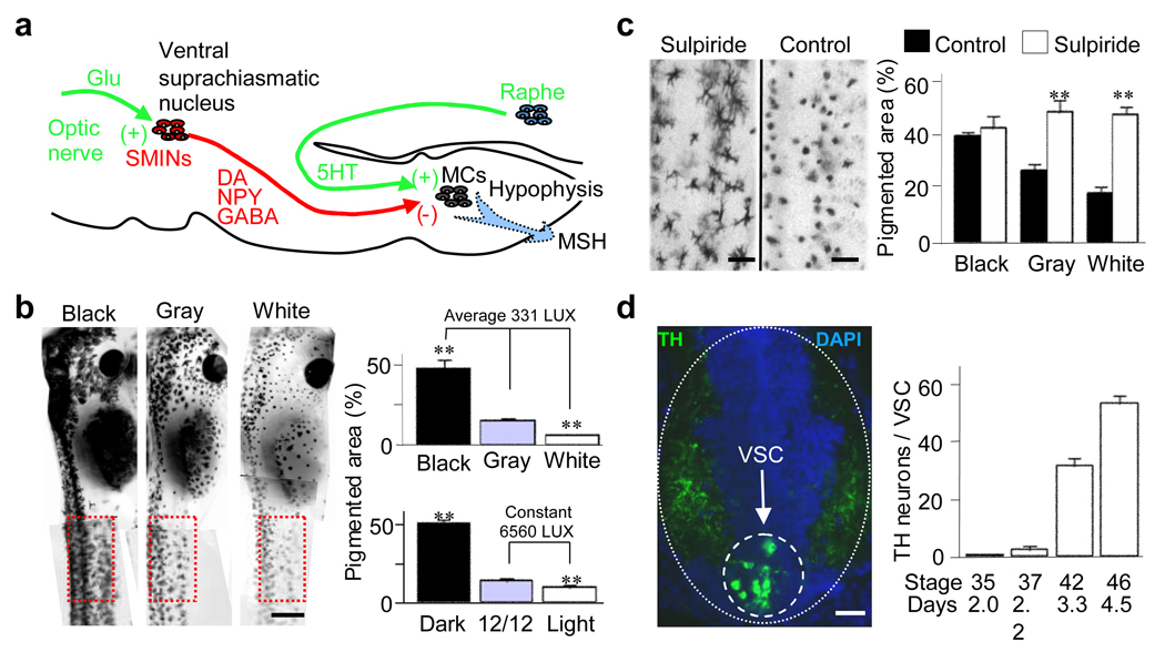
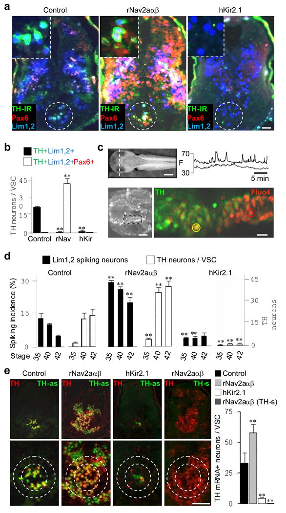
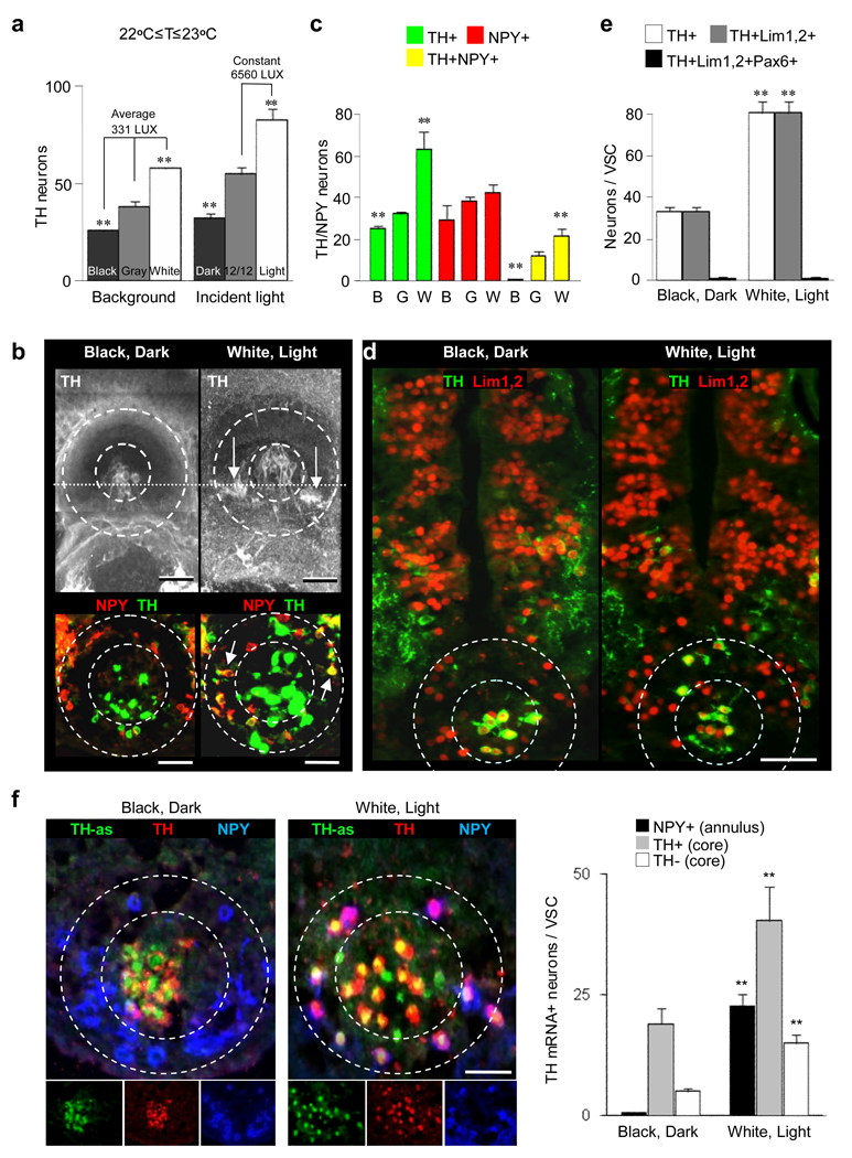
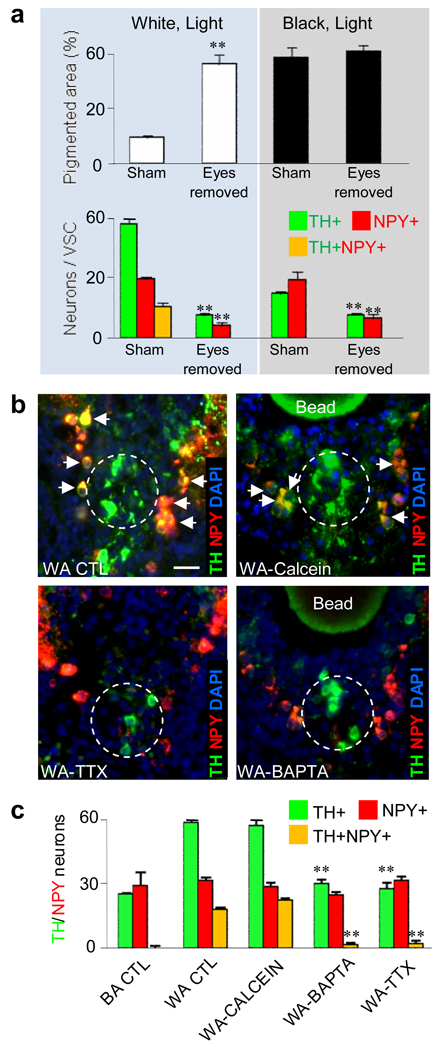
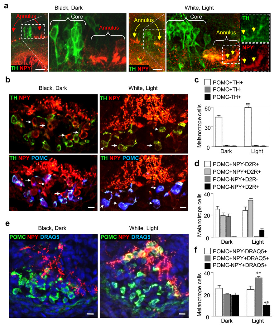
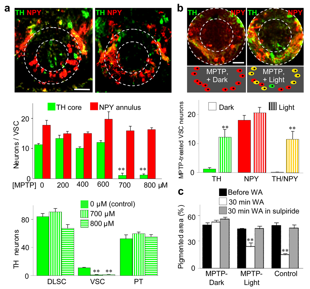
Specification of the appropriate neurotransmitter is a crucial step in neuronal differentiation because it enables signalling among populations of neurons. Experimental manipulations demonstrate that both autonomous and activity-dependent genetic programs contribute to this process during development, but whether natural environmental stimuli specify transmitter expression in a neuronal population is unknown. We investigated neurons of the ventral suprachiasmatic nucleus that regulate neuroendocrine pituitary function in response to light in teleosts, amphibia and primates. Here we show that altering light exposure, which changes the sensory input to the circuit controlling adaptation of skin pigmentation to background, changes the number of neurons expressing dopamine in larvae of the amphibian Xenopus laevis in a circuit-specific and activity-dependent manner. Neurons newly expressing dopamine then regulate changes in camouflage colouration in response to illumination. Thus, physiological activity alters the numbers of behaviourally relevant amine-transmitter-expressing neurons in the brain at postembryonic stages of development. The results may be pertinent to changes in cognitive states that are regulated by biogenic amines.


Figures

Figure 1
Figure 1
Dopaminergic VSC neurons regulate skin pigmentation. a, The diagram illustrates the neuronal circuit controlling this behavior. Glu, glutamate; SMINs, suprachiasmatic melanotrope inhibitory neurons; MC, melanotrope cells; MSH, melanocyte stimulating hormone. b, Raising animals in different levels of light changes pigmentation. A constant level of incident light was tested with different backgrounds and different levels of incident light were tested on a gray background. Illumination changes pigmentation: left, phenotypes on different backgrounds; right, pigmentation adaptation assayed in boxed regions at left. **, significantly different from intermediate illumination, p<0.001. N≥6 St 42 larvae for each condition. c, Activity of DA SMINs is necessary for regulation of pigmentation: left, 2 hr white adaptation in a larva treated with 10 nM sulpiride for 30 min compared to control; right, pigmentation at various background light levels in sulpiride-treated larvae compared to controls. **, significantly different, p<0.001. N≥6 St 42 larvae raised in the dark for each condition. d, The number of VSC neurons increases during development of larvae raised 12 hr light/12 hr dark on a gray background: left, transverse section through the St 42 diencephalon (dotted oval) illustrating the VSC (dashed circle); right, appearance of TH neurons during development. N≥6 larvae for each stage. Scale bars: b, 1 mm; c, 200 µm; d, 50 µm.
Figure 2
Figure 2
Dopaminergic differentiation is activity-dependent. a, Molecular markers identify VSC neurons (dashed circles) in transverse sections in a control larva and in larvae following sodium or potassium channel overexpression (St 42). The VSC is shown at higher magnification in insets (top left). b, Altering spike activity drives proportional changes in the number of TH neurons. Quantification of data presented in (a). N≥6 larvae for each condition. **, significantly different from control, p<0.001. c, Neurons generate Ca spike activity in the developing brain: left, transverse section through the hypothalamus (dashed line) at stage 35 yields frontal view showing the Fluo-4AM-loaded tissue, below; confocal imaging, postfixation and staining are from the area in the inset. Right, digitized fluorescence (F) of representative cells circled in the panel below; traces are Ca spike activity recorded from dorsal (top trace, red circle) TH- and ventral (bottom trace, yellow circle) TH+ cells. N=3 larvae.d, Across developmental stages the number of TH neurons is inversely proportional to the incidence of spiking in Lim1,2 neurons in controls and following alterations of spike activity. **, significantly different from control, p<0.001. e, TH mRNA expression is altered by ion channel overexpression. VSC double labeling with TH antibodies and TH-antisense probe (TH-as) in transverse brain sections (top row) and enlargements of the VSC (dashed circles, bottom row) in larvae injected with cascade blue (left panel, control), overexpressing sodium channels (second panel), or potassium channels (third panel). Antisense probe binding in sodium channel-overexpressing larvae (probe-s, fourth panel). Quantification of the results of channel overexpression is shown at right. **, significantly different from control, p<0.001. a–e, Larvae were raised on a 12/12 day/night cycle on a gray background. N≥6 larvae for each condition. Scale bars: a, 50 µm; c, 500 µm (left), 30 µm (right); e, 100 µm.
Figure 3
Figure 3
Illumination changes the numbers of dopaminergic neurons selectively in the VSC. a, Raising animals in different levels of light changes the number of TH neurons in the VSC. and A constant level of incident light was tested with different backgrounds and different levels of incident light were tested on a gray background. **, indicates significantly different from intermediate illumination, p<0.001. N≥9 St 42 larvae for each condition. b, TH viewed in wholemounts from above (top) and in transverse sections (bottom) shows the core (dashed inner circle) and the annular DA neurons (arrows, between dashed circles) in 2 hr black- and white-adapted St 42 larvae. Dotted line in wholemounts indicates section orientation. c, Two hr exposure of larvae to different background illumination (B, black; G, gray; W, white) changes the number of TH/NPY neurons in the VSC (core + annulus). ** indicates significantly different from gray background, p<0.001. d, Core DA neurons and newly recruited annular DA neurons from 2 hr white-adapted larvae are both from the Lim1,2 pool; sections as in (b). e, Exposure to different background illumination for 2 hr changes the number of Lim1,2 neurons expressing TH. ** indicates significantly different from dark rearing, p<0.001. f, TH mRNA expression is altered by light or dark adaptation. Triple labeling of transverse VSC sections (dashed circles) with TH, NPY and TH-as probe in 2 hr dark-, black-adapted and 2 hr light-, white-adapted larvae. Merged images (top row) and separate channels (bottom row) are shown. Quantification of the results of differential illumination is presented at right. **, value following light adaptation is significantly different from value following dark adaptation, p<0.001. b–f, St 42 larvae raised in the dark. c,e,f, N≥6 larvae for each condition. Scale bars: b, 100 µm (top), 60 µm (bottom); d, 100 µm; f, 60 µm.
Figure 4
Figure 4
Blocking physiological activity blocks illumination-dependent changes in numbers of TH VSC neurons. a, Binocular eye enucleation abolishes white adaptation of skin pigmentation and background illumination-dependent changes in numbers of DA neurons. **, significantly different relative to sham operation, p<0.001. b, Implanted beads delivering activity blockers prevent appearance of TH/NPY neurons in larvae exposed to 2 hr light relative to wild type or diffusion marker controls. Transverse sections through the VSC (dashed circles) show NPY/TH colocalization (yellow, arrows) in white-adapted wild type (WA WT) and calcein bead-implanted larvae that is absent in TTX and BAPTA-bead implanted larvae. Scale bar: 40 µm. c, TH and NPY identify VSC neurons in WA WT and BA WT (black-adapted wild type) controls and following suppression of activity. **, significantly different relative to calcein-bead implanted larvae, p<0.001. a,c, N≥6 St 42 larvae raised in the dark for each condition.
Figure 5
Figure 5
NPY neurons projecting to melanotrope cells express TH following illumination of larvae. a, Left, TH is absent in NPY annular neurons (red arrows) in transverse sections of larvae dark-adapted for 2 hr. Right, TH is present in somata (yellow arrows) and axons (yellow arrowheads) of NPY annular neurons in 2 hr white- and light-adapted larvae; color separation of green TH and red NPY (insets far right) facilitates visualization of double labeling of axons. b, Left, TH nerve terminals invest POMC-stained melanotrope cells in larvae dark-adapted for 2 hr, and terminals that are NPY and not TH project to melanotrope cells in which POMC is not detected (arrows). Right, TH/NPY terminals project to POMC melanotrope cells (arrows) in 2 hr white-adapted larvae. c, Quantification of the changes illustrated in (b). **, significantly different from value from dark-adapted larvae, p<0.001. d, D2 receptors are expressed in melanotrope cells of larvae light-adapted for 2 hr that newly express or may be acquiring POMC (light gray and black bars). e, DRAQ5-labeled nuclei identify 2 hr dark/light-dependent changes in melanotrope POMC expression f, Quantification of the changes illustrated in (e). **, significantly different from dark-adapted values, p<0.001. a–f, larvae raised in the dark. c,d,f, N≥6 stage 42 larvae for each condition. Scale bars: a, 100 µm; b, e, 40 µm.
Figure 6
Figure 6
Newly DA neurons regulate pigmentation. a, MPTP selectively eliminates the core VSC neurons. Top, TH/NPY in transverse sections before (left) and after (right) 8 hr incubation of larvae in 700 µM MPTP in the dark. Middle, the effect of different MPTP concentrations on the number of TH/NPY VSC neurons. Bottom, 700 µM MPTP eliminates VSC but not dorsolateral suprachiasmatic nucleus (DLSC) and posterior tuberculum (PT) DA nuclei. **, significantly different from 0 µM MPTP, p<0.001. b, Top, following MPTP treatment TH is largely absent from NPY annular neurons after dark-adaptation (left), and abundant following 2 hr light-adaptation (right). Middle, scheme illustrating transmitter expression in annular neurons in these conditions. Bottom, quantification of induction of TH; **, significantly different from values from dark adapted larvae, p<0.001. a,b, N>6 St 42 larvae raised in the dark. Scale bars: 50 µm. c, Illumination-dependent changes in skin pigmentation are rescued when MPTP treatment is followed by 2 hr exposure to light. Pigmentation of three groups of larvae raised in the dark: 1) MPTP-treated in the dark and kept in the dark; 2) MPTP-treated in the dark followed by 2 hr exposure to light; or 3) MPTP-untreated followed by 2 hr exposure to light (control). Pigmentation was measured before (black bars) or after (white bars) 30 min of white adaptation (WA), or after WA in the presence of sulpiride (gray bars). 3 left bars: MPTP followed by 2 hr incubation in the dark abolishes the change in pigmentation in response to 30 min white background adaptation. 3 center bars: MPTP followed by 2 hr incubation in the light recovers most of the change in pigmentation in response to a 30 min exposure to white background, relative to control (3 right bars). Sulpiride (10 nM) during 30 min exposure to white background blocks the reduction in pigmented area (gray bars, center and right groups). **, significantly different from value prior to white adaptation, p<0.001. N≥10 St 42 larvae.

Comment in

References

    1. Walicke PA, Patterson PH. On the role of Ca2+ in the transmitter choice made by cultured sympathetic neurons. Journal of Neuroscience. 1981;1:343–350. - PMC - PubMed
    1. Brosenitsch TA, Katz DM. Expression of Phox2 transcription factors and induction of the dopaminergic phenotype in primary sensory neurons. Molecular and Cellular Neuroscience. 2002;20:447–457. - PubMed
    1. Borodinsky LN, et al. Activity-dependent homeostatic specification of transmitter expression in embryonic neurons. Nature. 2004;429:523–530. - PubMed
    1. Gomez-Lira G, Lamas M, Romo-Parra H, Gutierrez R. Programmed and induced phenotype of the hippocampal granule cells. Journal of Neuroscience. 2005;25:6939–6946. - PMC - PubMed
    1. Catalano SM, Chang CK, Shatz CJ. Activity-dependent regulation of NMDAR1 immunoreactivity in the developing visual cortex. Journal of Neuroscience. 1997;17:8376–8390. - PMC - PubMed

Publication types

MeSH terms

Substances