. 2008 Nov 13;456(7219):202-8.
doi: 10.1038/nature07473.

Deconstructing voltage sensor function and pharmacology in sodium channels

Affiliations

Deconstructing voltage sensor function and pharmacology in sodium channels

Frank Bosmans et al. Nature. .

Abstract

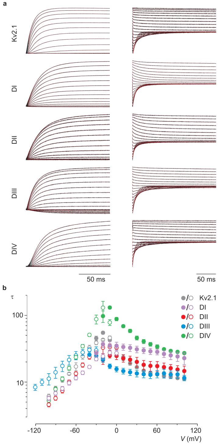
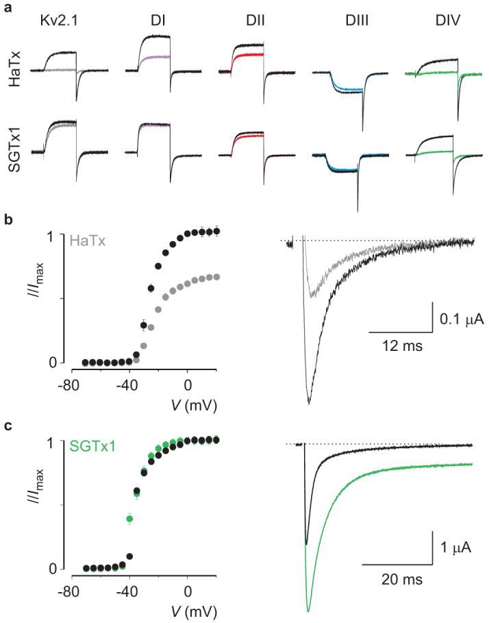
Voltage-activated sodium (Na(v)) channels are crucial for the generation and propagation of nerve impulses, and as such are widely targeted by toxins and drugs. The four voltage sensors in Na(v) channels have distinct amino acid sequences, raising fundamental questions about their relative contributions to the function and pharmacology of the channel. Here we use four-fold symmetric voltage-activated potassium (K(v)) channels as reporters to examine the contributions of individual S3b-S4 paddle motifs within Na(v) channel voltage sensors to the kinetics of voltage sensor activation and to forming toxin receptors. Our results uncover binding sites for toxins from tarantula and scorpion venom on each of the four paddle motifs in Na(v) channels, and reveal how paddle-specific interactions can be used to reshape Na(v) channel activity. One paddle motif is unique in that it slows voltage sensor activation, and toxins selectively targeting this motif impede Na(v) channel inactivation. This reporter approach and the principles that emerge will be useful in developing new drugs for treating pain and Na(v) channelopathies.


Figures

Figure 1
Figure 1. Transfer of the voltage sensor paddle motifs from rNav1.2a to Kv2.1
a, Cartoon depicting the paddle motif transfer from the Nav channel S1-S4 voltage sensor of domain II into Kv2.1. Purple: domain I paddle (DI), red: domain II paddle (DII), blue: domain III paddle (DIII) and green: domain IV paddle (DIV). Color code will be used in all figures. b,c, Families of potassium currents (b) and tail current voltage-activation relationships (c) for each chimaera (n = 18, error bars represent s.e.m.). Holding voltage was -90mV and the tail voltage was -50mV (-80mV for DIII). Scale bars in (b) are 2 μA and 100 ms.
Figure 2
Figure 2. Sensitivity of rNav1.2a paddle chimaeras to extracellular toxins
a, Effects of toxins on Kv2.1 and chimaeras where paddle motifs were transferred from rNav1.2a into Kv2.1. Normalized tail current voltage-activation relationships are shown where tail current amplitude is plotted against test voltage before (black) and in the presence of toxins (other colors). Data are grouped per toxin (horizontally) and per chimaera or wild-type Kv2.1 (vertically). Concentrations used are 100nM PaurTx3, ProTx-I and ProTx-II; 1μM AaHII; 500nM TsVII and 100μM VTD. b, Effects of TsVII (50 nM) on rNav1.4 paddle chimaeras. n = 3-5 and error bars represent s.e.m. The plant alkaloid veratridine (VTD) is used as a negative control. The holding voltage was -90mV, test pulse duration was 300 ms, and the tail voltage was -50mV (-80mV for DIII).
Figure 3
Figure 3. Scanning mutagenesis of Nav channel paddle motifs
a-d, Ala-scan of the separate rNav1.2a paddle motifs in the DI to DIV chimaeras where changes in apparent toxin affinity (Kdmut/Kdcont) are plotted for individual mutants. See Full Methods, Supplementary Fig. 4, 5c and Supplementary Table 3 for information on Kd measurements. Most of the residues within the rNav1.2a paddle in the four chimaera constructs were individually mutated to Ala (except for native Ala residues, which were mutated to Val). The dashed line marks a value of 1. Each mutant was initially examined using a concentration near the Kd value determined for the control chimaera (see Supplementary Fig. 4). Mutants with a Kdmut/Kdcont value greater than five were further examined using a wider range of concentrations. Glu residues marked with asterisks were also mutated to a Lys. Bar diagrams are approximately aligned according to the sequence alignment of the different paddles (see Supplementary Fig. 1). Mutants without a corresponding bar did not result in functional channels. Residues with a grey background were used in subsequent tests (Fig. 4). Mutation of two underlined residues in (d) results in an increase in AaHII affinity (Kd = 235 ± 24nM for R1629A, 205 ± 23nM for L1630A and 1902 ± 102nM for the DIV chimera). n = 3-5 for each toxin concentration and error bars represent s.e.m.
Figure 4
Figure 4. Reconstitution of paddle mutants into rNav1.2a and their effects on toxin-channel interactions
a, Concentration-dependence for ProTx-II inhibition of rNav1.2a and selected mutants plotted as fraction unbound (Fu) measured at negative voltages. Solid lines are fits of the Hill equation to the data with apparent Kd values shown. See Supplementary Fig. 5b and Supplementary Table 3 for further information. b, Normalized conductance-voltage relationships for rNav1.2a and two DIII mutants before and after addition of 50nM TsVII. Arrow indicates toxin effect or lack thereof. Holding voltage was -90 mV. c, rNav1.2a mutation L1630A increases affinity for AaHII. Sodium currents were elicited by a depolarization to -15 mV from a holding voltage of -90 mV. Green trace is after AaHII addition.
Figure 5
Figure 5. Kinetics of opening and closing for rNav1.2a/Kv2.1 chimaeras
a, Representative macroscopic currents (black) showing channel activation (left) and channel deactivation (right) using the following voltage protocols: activation, 10 mV incrementing steps to voltages between -40mV and +100mV from a holding potential of -90 mV; deactivation, 10 mV incrementing steps to voltages between 0mV and -100mV (-120mV for DIII) from a test voltage of between +80 and +100 mV (holding potential is -90 mV). Superimposed red curves are single exponential fits to the current records after initial lags in current activation. b, Mean time constants (τ) from single exponential fits to channel activation (filled circles) and deactivation (open circles) plotted as a function of the voltage at which the current was recorded. n = 4-8 and error bars represent s.e.m.
Figure 6
Figure 6. Identifying a tarantula toxin selective for the paddle motif in domain IV
a, Potassium currents elicited by depolarizations near the foot of the voltage-activation curve for Kv2.1 and chimaeras in the absence and presence of 50nM HaTx or 100nM SGTx1. The holding voltage was -90mV, test pulse duration was 300 ms, and the tail voltage was -50mV (-80mV for DIII). b, Conductance-voltage relationships for rNav1.2a before and after addition of 50nM HaTx (left), normalized to the maximal conductance in control. Sodium current elicited by a depolarization to -30 mV before and after addition of 50nM HaTx (right). c, Conductance-voltage relationship of rNav1.2a before and after addition of 100nM SGTx1 (left), individually normalized to the maximal conductance in either control or the presence of toxin. Sodium current elicited by a depolarization to -15 mV before and after addition of 100nM SGTx1 (right). n = 3-5 for each toxin concentration and error bars represent s.e.m (b, c).

Comment in

Similar articles

Cited by

References

    1. Frech GC, VanDongen AM, Schuster G, Brown AM, Joho RH. A novel potassium channel with delayed rectifier properties isolated from rat brain by expression cloning. Nature. 1989;340:642–5. - PubMed
    1. Stuhmer W, et al. Molecular basis of functional diversity of voltage-gated potassium channels in mammalian brain. Embo J. 1989;8:3235–44. - PMC - PubMed
    1. Tempel BL, Papazian DM, Schwarz TL, Jan YN, Jan LY. Sequence of a probable potassium channel component encoded at Shaker locus of Drosophila. Science. 1987;237:770–5. - PubMed
    1. Auld VJ, et al. A rat brain Na+ channel alpha subunit with novel gating properties. Neuron. 1988;1:449–61. - PubMed
    1. Trimmer JS, et al. Primary structure and functional expression of a mammalian skeletal muscle sodium channel. Neuron. 1989;3:33–49. - PubMed

Publication types

MeSH terms