Inhibition of expression in Escherichia coli of a virulence regulator MglB of Francisella tularensis using external guide sequence technology
- PMID: 19005569
- PMCID: PMC2579583
- DOI: 10.1371/journal.pone.0003719
Inhibition of expression in Escherichia coli of a virulence regulator MglB of Francisella tularensis using external guide sequence technology
Abstract
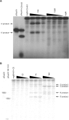
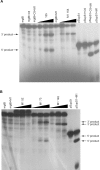
External guide sequences (EGSs) have successfully been used to inhibit expression of target genes at the post-transcriptional level in both prokaryotes and eukaryotes. We previously reported that EGS accessible and cleavable sites in the target RNAs can rapidly be identified by screening random EGS (rEGS) libraries. Here the method of screening rEGS libraries and a partial RNase T1 digestion assay were used to identify sites accessible to EGSs in the mRNA of a global virulence regulator MglB from Francisella tularensis, a Gram-negative pathogenic bacterium. Specific EGSs were subsequently designed and their activities in terms of the cleavage of mglB mRNA by RNase P were tested in vitro and in vivo. EGS73, EGS148, and EGS155 in both stem and M1 EGS constructs induced mglB mRNA cleavage in vitro. Expression of stem EGS73 and EGS155 in Escherichia coli resulted in significant reduction of the mglB mRNA level coded for the F. tularensis mglB gene inserted in those cells.
Conflict of interest statement
Figures


Similar articles
-
Targeted cleavage of mRNA in vitro by RNase P from Escherichia coli.Proc Natl Acad Sci U S A. 1992 Apr 15;89(8):3185-9. doi: 10.1073/pnas.89.8.3185. Proc Natl Acad Sci U S A. 1992. PMID: 1373488 Free PMC article.
-
Rapid selection of accessible and cleavable sites in RNA by Escherichia coli RNase P and random external guide sequences.Proc Natl Acad Sci U S A. 2008 Feb 19;105(7):2354-7. doi: 10.1073/pnas.0711977105. Epub 2008 Feb 8. Proc Natl Acad Sci U S A. 2008. PMID: 18263737 Free PMC article.
-
Artificial regulation of gene expression in Escherichia coli by RNase P.Proc Natl Acad Sci U S A. 1995 Nov 21;92(24):11115-9. doi: 10.1073/pnas.92.24.11115. Proc Natl Acad Sci U S A. 1995. PMID: 7479948 Free PMC article.
-
Loops and networks in control of Francisella tularensis virulence.Future Microbiol. 2009 Aug;4(6):713-29. doi: 10.2217/fmb.09.37. Future Microbiol. 2009. PMID: 19659427 Review.
-
The unraveling panoply of Francisella tularensis virulence attributes.Curr Opin Microbiol. 2010 Feb;13(1):11-7. doi: 10.1016/j.mib.2009.11.007. Epub 2009 Dec 23. Curr Opin Microbiol. 2010. PMID: 20034843 Review.
Cited by
-
RNase P-Mediated Sequence-Specific Cleavage of RNA by Engineered External Guide Sequences.Biomolecules. 2015 Nov 9;5(4):3029-50. doi: 10.3390/biom5043029. Biomolecules. 2015. PMID: 26569326 Free PMC article. Review.
-
Inhibition of cell division induced by external guide sequences (EGS Technology) targeting ftsZ.PLoS One. 2012;7(10):e47690. doi: 10.1371/journal.pone.0047690. Epub 2012 Oct 23. PLoS One. 2012. PMID: 23110089 Free PMC article.
-
Inactivation of expression of two genes in Saccharomyces cerevisiae with the external guide sequence methodology.RNA. 2011 Mar;17(3):544-9. doi: 10.1261/rna.2538711. Epub 2011 Jan 13. RNA. 2011. PMID: 21233222 Free PMC article.
-
External guide sequence technology: a path to development of novel antimicrobial therapeutics.Ann N Y Acad Sci. 2015 Sep;1354(1):98-110. doi: 10.1111/nyas.12755. Epub 2015 Apr 9. Ann N Y Acad Sci. 2015. PMID: 25866265 Free PMC article. Review.
-
Inactivating Gene Expression with Antisense Modified Oligonucleotides.Acta Naturae. 2021 Jul-Sep;13(3):101-105. doi: 10.32607/actanaturae.11522. Acta Naturae. 2021. PMID: 34707901 Free PMC article.
References
-
- Tärnvik A. Nature of protective immunity to Francisella tularensis. Rev Infect Dis. 1989;11:440–451. - PubMed
-
- Baron GS, Nano FE. MglA and MglB are required for the intramacrophage growth of Francisella novicida. Mol Microbiol. 1998;29:247–259. - PubMed
-
- Gray CG, Cowley SC, Cheung KK, Nano FE. The identification of five genetic loci of Francisella novicida associated with intracellular growth. FEMS Microbiol Lett. 2002;215:53–56. - PubMed
Publication types
MeSH terms
Substances
LinkOut - more resources
Full Text Sources
Molecular Biology Databases

