. 2008:14:2042-55.
Epub 2008 Nov 12.

The EPHA2 gene is associated with cataracts linked to chromosome 1p

Affiliations

The EPHA2 gene is associated with cataracts linked to chromosome 1p

Alan Shiels et al. Mol Vis. 2008.

Abstract

Purpose: Cataracts are a clinically and genetically heterogeneous disorder affecting the ocular lens, and the leading cause of treatable vision loss and blindness worldwide. Here we identify a novel gene linked with a rare autosomal dominant form of childhood cataracts segregating in a four generation pedigree, and further show that this gene is likely associated with much more common forms of age-related cataracts in a case-control cohort.

Methods: Genomic DNA was prepared from blood leukocytes, and genotyping was performed by means of single nucleotide polymorphism (SNP) markers, and short tandem repeat (STR) markers. Linkage analyses were performed with the GeneHunter and MLINK programs, and association analyses were performed with the Haploview and Exemplar programs. Mutation detection was achieved by PCR amplification of exons and di-deoxy cycle-sequencing.

Results: Genome-wide linkage analysis with SNP markers, identified a likely disease-haplotype interval on chromosome 1p (rs707455-[approximately 10 Mb]-rs477558). Linkage to chromosome 1p was confirmed using STR markers D1S2672 (LOD score [Z]=3.56, recombination distance [theta]=0), and D1S2697 (Z=2.92, theta=0). Mutation profiling of positional-candidate genes detected a heterozygous transversion (c.2842G>T) in exon 17 of the gene coding for Eph-receptor type-A2 (EPHA2) that cosegregated with the disease. This missense change was predicted to result in the non-conservative substitution of a tryptophan residue for a phylogenetically conserved glycine residue at codon 948 (p.G948W), within a conserved cytoplasmic domain of the receptor. Candidate gene association analysis further identified SNPs in the EPHA2 region of chromosome 1p that were suggestively associated with age-related cataracts (p=0.007 for cortical cataracts, and p=0.01 for cortical and/or nuclear cataracts).

Conclusions: These data provide the first evidence that EPHA2, which functions in the Eph-ephrin bidirectional signaling pathway of mammalian cells, plays a vital role in maintaining lens transparency.


Figures

Figure 1
Figure 1
Autosomal dominant posterior polar cataracts in a four generation white American pedigree (family Mu). A: Pedigree and haplotype analysis showing segregation of seven STR markers on chromosome 1p listed in descending order from the telomere. Squares and circles denote males and females, respectively. Filled symbols and bars denote affected status and haplotypes, respectively. B: Slit-lamp image of left lens from affected female IV:6 (age 12 years) showing posterior sub-capsular opacity. C: Ideogram of chromosome 1p36, comparing the cytogenetic and physical locations of STR markers defining the posterior polar cataract locus in this study (red) with those defining 3 other loci (black) for autosomal dominant cataracts (CCV, CTPP1 and Total) [45-47], and a locus (blue) for age-related cortical cataracts [44]. M, mega-base pairs.
Figure 2
Figure 2
Parametric multipoint LOD scores (pLOD) for linkage between the posterior polar cataract phenotype in family Mu and SNP markers across the 22 autosomes.
Figure 3
Figure 3
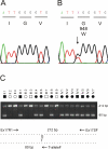
Mutation analysis of EPHA2 in family Mu. A: Sequence trace of the wild type allele showing translation of glycine (G) at codon 948 (GGG). B: Sequence trace of the mutant allele showing the heterozygous c.2842G>T transversion (denoted K by the International Union of Pure and Applied Chemistry [IUPAC] code) that is predicted to result in the missense substitution of tryptophan (TGG) for glycine at codon 948 (p.G948W). C: Allele-specific PCR analysis using the 3 primers (Table 1) indicated by arrows in the schematic diagram; exon-17 was amplified as above with the sense (anchor) primer located in intron 16 (Ex17R1), the anti-sense primer located in the 3′-untranslated region (Ex17SF), and the nested mutant primer specific for the T-allele in codon 948 (T-alleleF). Note that only affected members of family Mu are heterozygous for the mutant T-allele (80 bp).
Figure 4
Figure 4
Schematic diagram showing gene structure and protein domains of EPHA2. A: Exon-intron organization, mutation profile and protein domains. Intron sizes are shown in base-pairs (bp), and codons are numbered below each exon (boxes). The approximate locations of protein domains are indicated. SP, signal-peptide; Eph-lbd, Eph-receptor ligand binding domain; EGF-like, epithelial growth factor-like region; Fn-3, fibronectin type-III domain; Tm-1, transmembrane domain type-1; Pkinase-Tyr, protein tyrosine kinase domain; SAM, sterile-α-motif. The predicted p.G948W missense mutation identified in family Mu is proposed to reside in the cytoplasmic SAM domain. B: Amino acid sequence alignment of the SAM domain from human EPHA2 and orthologs from other species, showing conservation of p.G948. Divergent amino-acids residues are shaded. The SAM domain comprises an NH2-terminal extended strand (dots), 5 α-helices (H1–5, over-lined), and a core sub-unit (bolded). The proposed p.G948W substitution likely resides between H-4 and H-5.
Figure 5
Figure 5
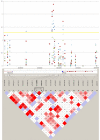
Graphical presentation of association between SNPs in the EPHA2 region of chromosome 1p with age-related cataracts in the Italian case-control cohort. The upper panel shows a plot of -log p-values (y-axis) from association analyses of 21 SNPs across the EPHA2 region. Blue diamonds denote -log p values for pure cortical cataracts; red squares, any cortical cataracts; green triangles, pure nuclear cataracts; purple x, any nuclear cataracts; and light blue asterisks, any cataracts. The x-axis shows the relative physical location of each SNP measured in mega-base-pairs. The lower panel shows a pairwise linkage disequilibrium (D´) Haploview plot for SNPs in the EPHA2 region. The strength of linkage disequilibrium (LD) is color-coded; red indicates strong LD with SNPs showing high correlation, and blue indicates low LD and high recombination. The relative positions of EPHA2 and the adjacent FAM131C are indicated with haplotype blocks for the European population (CEU) from the HapMap project (SNPbrowser, Applied Biosystems).

Similar articles

Cited by

References

    1. World Health Organization - Vision 2020; The Right to Sight, Global initiative for the elimination of avoidable blindness - Action plan 2006–2011 (http://www.who.int/blindness/Vision2020%20-report.pdf)
    1. SanGiovanni JP. Infantile cataract in the collaborative perinatal project: prevalence and risk factors. Arch Ophthalmol. 2002;120:1559–65. - PubMed
    1. Zetterstrom C. Cataracts in children. J Cataract Refract Surg. 2005;31:824–40. - PubMed
    1. Shiels A. Genetic origins of cataract. Arch Ophthalmol. 2007;125:165–73. - PubMed
    1. Litt M. Autosomal dominant congenital cataract associated with a missense mutation in the human alpha crystallin gene CRYAA. Hum Mol Genet. 1998;7:471–4. - PubMed

Publication types

MeSH terms

Substances

LinkOut - more resources