

A three-dimensional retrospective analysis of the evolution of spinal instrumentation for the correction of adolescent idiopathic scoliosis
- PMID: 19005692
- PMCID: PMC2615118
- DOI: 10.1007/s00586-008-0817-4
A three-dimensional retrospective analysis of the evolution of spinal instrumentation for the correction of adolescent idiopathic scoliosis
Abstract
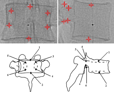
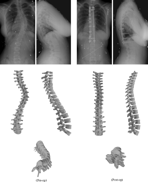
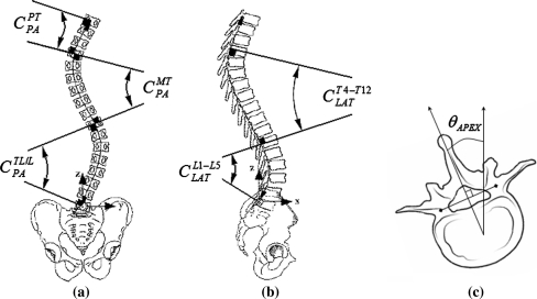
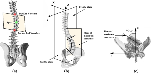
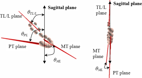
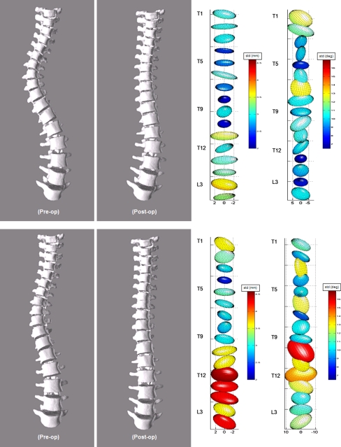
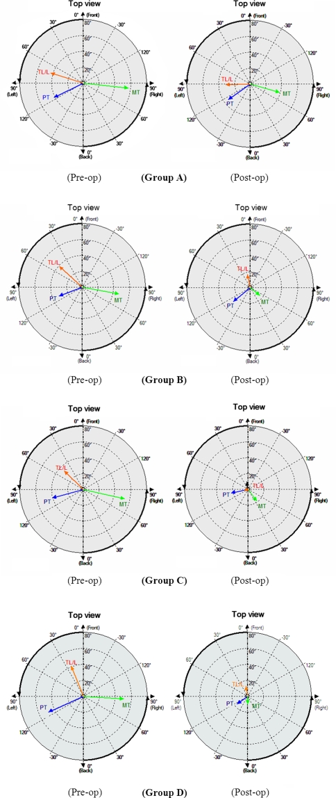
This is a clinical radiographic study, spanning over three decades, analyzing the three-dimensional (3-D) changes in spine geometry after corrective surgery for adolescent idiopathic scoliosis (AIS) using four generations of instrumentation systems. The objective of this study was to retrospectively evaluate the evolution of spinal instrumentation over time by measuring the 3-D changes of spinal shape before and after surgical correction of subjects with AIS using Harrington/Harrington-Luque (H/HL) instrumentation, original and recent generations of Cotrel-Dubousset Instrumentation (CDI) with rod rotation maneuvers, as well as third generation systems using thoracic pedicle screws and direct vertebral derotation (DVD) manoeuver in order to determine if the claims for improved 3-D correction from generation to next generation could be substantiated. The 3-D shape of the thoracic and lumbar spine was recorded from a pair of standing radiographs using a novel 3-D reconstruction technique from uncalibrated radiographs in 128 adolescents with AIS undergoing surgery by a posterior approach. Changes in coronal Cobb angles, kyphosis, lordosis, as well as in a series of 3-D parameters computed from the spine reconstructions before and after surgery were used to compare the four groups. Results demonstrate statistically significant differences (P = 0.05) between generations with regards to the correction of the coronal Cobb angle, and different loss of physiological lordosis. More importantly, significant differences in the 3-D correction of the spine based on the orientation of the planes of maximal curvature were observed (20/-6% H/HL vs. 39/39% CDI vs. 42/18% DVD for the thoracic/lumbar regions, respectively), confirming that recent CDI and third generation instrumentations coupled with DVD can bring the deformity significantly closer to the sagittal plane. An increased correction in apical vertebra axial rotation was observed with the DVD manoeuver (74%), while fewer notable differences were found between DVD and recent CDI systems in terms of 3-D correction. This is the first quantitative study to clearly demonstrate that the rod derotation and DVD maneuvers can significantly improve 3-D correction of scoliotic deformities, thereby supporting the transition towards these more elaborate and costly instrumentation technologies in terms of 3-D assessment.
Figures


References
-
- Aubin C-E, Lobeau D, Labelle H, Maquinghen-Godillon AP, LeBlanc R, Dansereau J (1999) Planes of Maximum Deformity in the Scoliotic Spine. In: IAF Stokes (ed) Research into spinal deformities 2. IOS Press, Amsterdam, pp 45–48
-
- Bassett GS, Hensinger MC, Keiper MD (1989) Effect of posterior spinal fusion on spinal balance in idiopathic scoliosis. J Pediatr Orthop 9:672–674 - PubMed
Publication types
MeSH terms
LinkOut - more resources
Full Text Sources
Medical
