

Anatomical study of axis for odontoid screw thickness, length, and angle
- PMID: 19005694
- PMCID: PMC2899344
- DOI: 10.1007/s00586-008-0814-7
Anatomical study of axis for odontoid screw thickness, length, and angle
Abstract
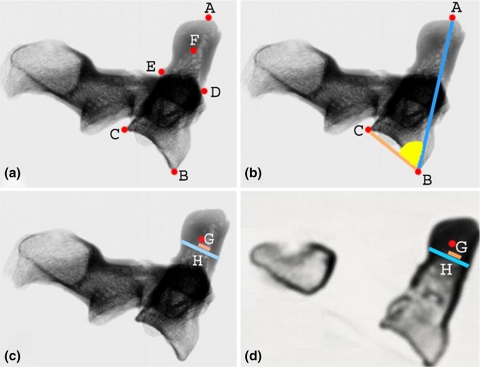
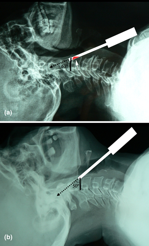
Anterior odontoid screw fixation is a safe and effective method for treatment of odontoid fractures. The screw treads should fit into the odontoid medulla, should pass the fracture line, and should pull fractured odontoid tip against body of axis in order to achieve optimum screw placement and treatment. This study has demonstrated optimal anterior odontoid screw thickness, length, and optimal angle for safe and strong anterior odontoid screw placement. Dry bone axis vertebrae were evaluated by direct measurements, X-ray measurements, and computerized tomography (CT) measurements. The screw thickness (inner diameter of the odontoid) was measured as well as screw length (distance between anterior-inferior point body of axis and tip of odontoid), and screw angle (the angle between basis of axis and tip of odontoid). The inner diameter of odontoid bone was measured as 6.5+/-1.9 mm, the screw length was 37.6+/-3.3 mm, and the screw angle was 62.4+/-4.7 on CT. There was no statistical difference between X-ray and CT in the measurements of screw thickness and angle. X-ray and CT measurements are both safe methods to determine the inner odontoid diameter and angle preoperatively. Screw length should be measured on CT only. To provide safe and strong anterior odontoid screw fixation, screw thickness, length, and angle should be known preoperatively, and these can be measured on X-ray and CT.
Figures
References
-
- Alfieri A. Single-screw fixation for acute type II odontoid fracture. J Neurosurg Sci. 2001;45:15–18. - PubMed
-
- Anderson LD, D’Alonzo RT. Fractures of the odontoid process of the axis. J Bone Joint Surg Am. 1974;56:1663–1674. - PubMed
-
- Carlson GD, Heller JG, Abitbol JJ. Odontoid fractures. In: Levine AM, editor. Spine trauma. Philadelphia: WB Saunders Co; 1998. pp. 227–248.
MeSH terms
LinkOut - more resources
Full Text Sources
Medical
