

Cell transplantation in lumbar spine disc degeneration disease
- PMID: 19005697
- PMCID: PMC2587656
- DOI: 10.1007/s00586-008-0750-6
Cell transplantation in lumbar spine disc degeneration disease
Abstract
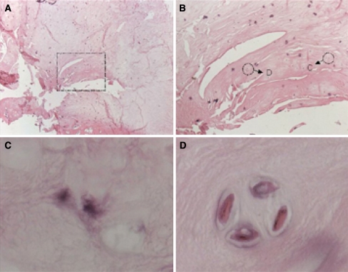
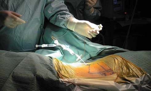
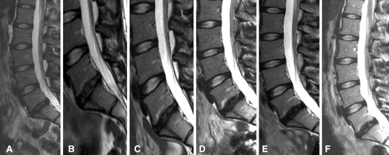
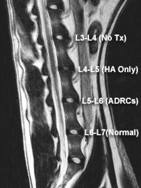
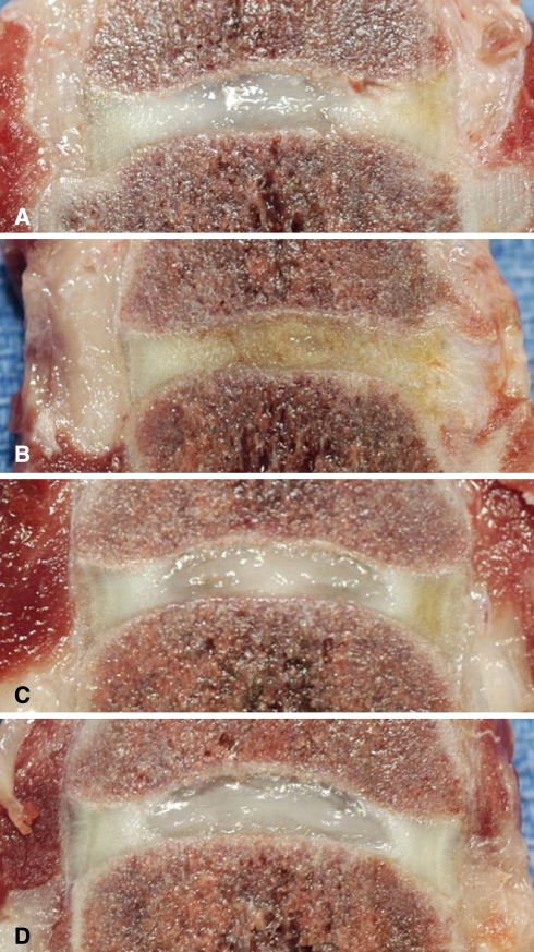
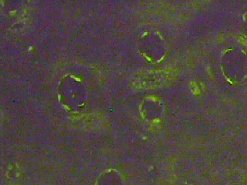
Low back pain is an extremely common symptom, affecting nearly three-quarters of the population sometime in their life. Given that disc herniation is thought to be an extension of progressive disc degeneration that attends the normal aging process, seeking an effective therapy that staves off disc degeneration has been considered a logical attempt to reduce back pain. The most apparent cellular and biochemical changes attributable to degeneration include a decrease in cell density in the disc that is accompanied by a reduction in synthesis of cartilage-specific extracellular matrix components. With this in mind, one therapeutic strategy would be to replace, regenerate, or augment the intervertebral disc cell population, with a goal of correcting matrix insufficiencies and restoring normal segment biomechanics. Biological restoration through the use of autologous disc chondrocyte transplantation offers a potential to achieve functional integration of disc metabolism and mechanics. We designed an animal study using the dog as our model to investigate this hypothesis by transplantation of autologous disc-derived chondrocytes into degenerated intervertebral discs. As a result we demonstrated that disc cells remained viable after transplantation; transplanted disc cells produced an extracellular matrix that contained components similar to normal intervertebral disc tissue; a statistically significant correlation between transplanting cells and retention of disc height could displayed. Following these results the Euro Disc Randomized Trial was initiated to embrace a representative patient group with persistent symptoms that had not responded to conservative treatment where an indication for surgical treatment was given. In the interim analyses we evaluated that patients who received autologous disc cell transplantation had greater pain reduction at 2 years compared with patients who did not receive cells following their discectomy surgery and discs in patients that received cells demonstrated a significant difference as a group in the fluid content of their treated disc when compared to control. Autologous disc-derived cell transplantation is technically feasible and biologically relevant to repairing disc damage and retarding disc degeneration. Adipose tissue provides an alternative source of regenerative cells with little donor site morbidity. These regenerative cells are able to differentiate into a nucleus pulposus-like phenotype when exposed to environmental factors similar to disc, and offer the inherent advantage of availability without the need for transporting, culturing, and expanding the cells. In an effort to develop a clinical option for cell placement and assess the response of the cells to the post-surgical milieu, adipose-derived cells were collected, concentrated, and transplanted under fluoroscopic guidance directly into a surgically damaged disc using our dog model. This study provides evidence that cells harvested from adipose tissue might offer a reliable source of regenerative potential capable of bio-restitution.
Figures

Similar articles
-
Clinical experience in cell-based therapeutics: disc chondrocyte transplantation A treatment for degenerated or damaged intervertebral disc.Biomol Eng. 2007 Feb;24(1):5-21. doi: 10.1016/j.bioeng.2006.07.002. Epub 2006 Jul 21. Biomol Eng. 2007. PMID: 16963315
-
A prospective multicenter phase I/II clinical trial to evaluate safety and efficacy of NOVOCART Disc plus autologous disc chondrocyte transplantation in the treatment of nucleotomized and degenerative lumbar disc to avoid secondary disease: study protocol for a randomized controlled trial.Trials. 2016 Feb 26;17(1):108. doi: 10.1186/s13063-016-1239-y. Trials. 2016. PMID: 26920137 Free PMC article. Clinical Trial.
-
Disc chondrocyte transplantation in a canine model: a treatment for degenerated or damaged intervertebral disc.Spine (Phila Pa 1976). 2003 Dec 1;28(23):2609-20. doi: 10.1097/01.BRS.0000097891.63063.78. Spine (Phila Pa 1976). 2003. PMID: 14652478
-
Tissue engineering and the intervertebral disc: the challenges.Eur Spine J. 2008 Dec;17 Suppl 4(Suppl 4):480-91. doi: 10.1007/s00586-008-0746-2. Epub 2008 Nov 13. Eur Spine J. 2008. PMID: 19005701 Free PMC article. Review.
-
Future perspectives of cell-based therapy for intervertebral disc disease.Eur Spine J. 2008 Dec;17 Suppl 4(Suppl 4):452-8. doi: 10.1007/s00586-008-0743-5. Epub 2008 Nov 13. Eur Spine J. 2008. PMID: 19005704 Free PMC article. Review.
Cited by
-
Edifying the Focal Factors Influencing Mesenchymal Stem Cells by the Microenvironment of Intervertebral Disc Degeneration in Low Back Pain.Pain Res Manag. 2022 Mar 27;2022:6235400. doi: 10.1155/2022/6235400. eCollection 2022. Pain Res Manag. 2022. PMID: 35386857 Free PMC article. Review.
-
Potential regenerative treatment strategies for intervertebral disc degeneration in dogs.BMC Vet Res. 2014 Jan 4;10:3. doi: 10.1186/1746-6148-10-3. BMC Vet Res. 2014. PMID: 24387033 Free PMC article. Review.
-
A minimally invasive rabbit model of progressive and reproducible disc degeneration confirmed by radiology, gene expression, and histology.J Korean Neurosurg Soc. 2013 Jun;53(6):323-30. doi: 10.3340/jkns.2013.53.6.323. Epub 2013 Jun 30. J Korean Neurosurg Soc. 2013. PMID: 24003365 Free PMC article.
-
Differentiation of human induced pluripotent stem cells into nucleus pulposus-like cells.Stem Cell Res Ther. 2018 Mar 9;9(1):61. doi: 10.1186/s13287-018-0797-1. Stem Cell Res Ther. 2018. PMID: 29523190 Free PMC article.
-
Stem Cells and Exosomes: New Therapies for Intervertebral Disc Degeneration.Cells. 2021 Aug 29;10(9):2241. doi: 10.3390/cells10092241. Cells. 2021. PMID: 34571890 Free PMC article. Review.
References
-
- Annunen S, Paassilta P, Lohiniva J, Perala M, Pihlajamaa T, Karppinen J, Tervonen O, Kroger H, Lahde S, Vanharanta H, Ryhanen L, Goring HH, Ott J, Prockop DJ, Ala-Kokko L. An allele of COL9A2 associated with intervertebral disc disease. Science. 1999;285:409–412. doi: 10.1126/science.285.5426.409. - DOI - PubMed
-
- Antoniou J, Steffen T, Nelson F, Winterbottom N, Hollander AP, Poole RA, Aebi M, Alini M. The human lumbar intervertebral disc: evidence for changes in the biosynthesis and denaturation of the extracellular matrix with growth, maturation, ageing, and degeneration. J Clin Invest. 1996;98:996–1003. doi: 10.1172/JCI118884. - DOI - PMC - PubMed
-
- Beard HK, Roberts S, O’Brien JP. Immunofluorescent staining for collagen and proteoglycan in normal and scoliotic intervertebral discs. J Bone Joint Surg Br. 1981;63B:529–534. - PubMed
Publication types
MeSH terms
LinkOut - more resources
Full Text Sources
Other Literature Sources
Medical
Research Materials
