Review
. 2008 Dec;17 Suppl 4(Suppl 4):441-51.
doi: 10.1007/s00586-008-0749-z. Epub 2008 Nov 13.

Biological repair of the degenerated intervertebral disc by the injection of growth factors

Affiliations
Review

Biological repair of the degenerated intervertebral disc by the injection of growth factors

Koichi Masuda. Eur Spine J. 2008 Dec.

Abstract

The homeostasis of intervertebral disc (IVD) tissues is accomplished through a complex and precise coordination of a variety of substances, including cytokines, growth factors, enzymes and enzyme inhibitors. Recent biological therapeutic strategies for disc degeneration have included attempts to up-regulate the production of key matrix proteins or to down-regulate the catabolic events induced by pro-inflammatory cytokines. Several approaches to deliver these therapeutic biologic agents have been proposed and tested in a preclinical setting. One of the most advanced biological therapeutic approaches to regenerate or repair a degenerated disc is the injection of a recombinant growth factor. Abundant evidence for the efficacy of growth factor injection therapy for the treatment of IVD degeneration can be found in preclinical animal studies. Recent data obtained from animal studies on changes in cytokine expression following growth factor injection illustrate the great potential for patients with chronic discogenic low back pain. The first clinical trial for growth factor injection has been initiated and the results of that study may prove the usefulness of growth factor injection for treating the symptoms of patients with degenerative disc diseases. The focus of this review article is the effects of an in vivo injection of growth factors on the biological repair of the degenerated intervertebral disc in animal models. The effects of growth factor injection on the symptoms of patients with low back pain, the therapeutic target of growth factor injection and the limitations of the efficacy of growth factor therapy are also reviewed. Further quantitative studies on the effect of growth factor injection on pain generation and the long term effects on the endplate and cell survival after an injection using large animals are needed. An international academic-industrial consortium addressing these aims, such as was achieved for osteoarthritis (The Osteoarthritis Initiative), may further the development of biological therapies for degenerative disc diseases.


Figures
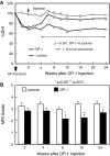
Fig. 1
Fig. 1
Changes in the intervertebral disc height index (DHI, a) and magnetic resonance imaging (MRI) grading (b) after anular puncture and osteogenic protein-1 (OP-1) injection (Modified from Masuda K et al. Osteogenic protein-1 injection into a degenerated disc induces the restoration of disc height and structural changes in the rabbit anular puncture model. Spine 2006;31(7):742–754). Adolescent New Zealand White rabbits (3–3.5 kg) received an anular puncture with an 18G needle to induce disc degeneration. Four weeks later, either 5% lactose (vehicle control; 10 μl) or osteogenic protein-1 (OP-1; 100 μg in 10 μl 5% lactose) was injected into the center of the nucleus pulposus (NP). By 4 weeks after the OP-1 injection, the mean %DHI of injected discs in the OP-1 group was significantly higher than that in the lactose control group (P < 0.001, repeated ANOVA). This significant difference in mean %DHI was maintained during the follow-up period (P < 0.001). MRI examinations were performed on all the spinal columns isolated from the rabbits ex vivo at the time of killing. When the degree of disc degeneration by MRI grading was compared between the lactose and OP-1 groups, a significant lower grading of MRI in the OP-1 group was observed (P < 0.01, Mann-Whitney U test)
Fig. 2
Fig. 2
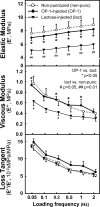
Effects of treatment with osteogenic protein-1 (OP-1) or lactose on the dynamic viscoelastic properties and the biochemical properties of rabbit intervertebral discs (IVDs) (Miyamoto K et al. Intradiscal injections of osteogenic protein-1 restore the viscoelastic properties of degenerated intervertebral discs. Spine J 2006;6(6):692–703). After killing and removal of bone-disc-bone complexes 8 weeks post-injection, the dynamic viscoelastic properties of the IVDs were tested by applying a cycle of sinusoidal strain in uniaxial compression at six loading frequencies (0.05–2 Hz). In the OP-1-injected discs (closed circles), at all loading frequencies, the elastic modulus (E′) was significantly higher than that in the lactose-injected discs and approached that of the non-punctured control discs (open circle) (OP-1 vs. non-punctured control, n.s., upper graph). The viscous modulus (E″) was significantly higher in the OP-1-injected discs (closed circle) than that in the lactose-injected discs (reverse triangle) (middle graph). No significant difference was observed in the loss tangent (E″/E′) among the non-punctured control and the rhOP-1-injected and lactose-injected discs
Fig. 3
Fig. 3
Effect of osteogenic protein-1 (OP-1) injection on the disc height after chemonucleolysis by chondroitinase-ABC (C-ABC) (With permission from Imai Y et al. Restoration of disc height loss by recombinant human osteogenic protein-1 injection into intervertebral discs undergoing degeneration induced by an intradiscal injection of chondroitinase ABC. Spine 2007;32:1197–205). Rabbit lumbar intervertebral discs (L2-5) were exposed through a posterolateral retroperitoneal approach and chondroitinase ABC (C-ABC) at 10 mU in 10 μl of saline was injected into the center of the nucleus pulposus of three consecutive intervertebral discs (L2/3, 3/4, 4/5). A significant decrease of disc height in the lateral radiograph was observed at 4 weeks. Four weeks after the initial injection of C-ABC, the vehicle (lactose, 10 μl) or rhOP-1 in lactose (100 μg/10 μl at each level) was injected. By 6 weeks after the rhOP-1 injection, the mean percent disc height index (%DHI) of the injected discs in the C-ABC/rhOP-1 group was significantly higher than in the C-ABC/lactose group (P < 0.01). This significant difference in mean %DHI was maintained during the follow-up period (P < 0.01)
Fig. 4
Fig. 4
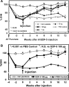
Changes in the intervertebral disc (IVD) height index (DHI) after anular puncture and recombinant human growth and differentiation factor-5 (rhGDF-5) injection (Modified from Chujo T et al. Effects of growth differentiation factor-5 on the intervertebral disc- in vitro bovine study and in vivo rabbit disc degeneration model study. Spine 2006;31(25):2909–2917 and Chujo T et al. In vivo effects of recombinant human growth and differentiation factor-5 on the repair of the mature rabbit intervertebral disc. Spine J, 6(5):23S–24S, 2006 (North American Spine Society, 21st Annual Meeting Proceeding, abstract). a Four weeks after inducing disc degeneration by an anular needle puncture in New Zealand White rabbits (weighing 3.5–4 kg), the rabbits received an injection of phosphate buffered saline (PBS) (10 μl) or rhGDF-5 (10 ng, 1 or 100 μg, in 10 μl PBS) and were monitored radiographically for up to 12 weeks after the injections. After killing, the specimens were assessed by magnetic resonance imaging (MRI) and histology. By 4 weeks after the rhGDF-5 injection, the mean %DHI of injected discs in the rhGDF-5 group (100 μg) was significantly higher than in the PBS group (P < 0.05). After 12 weeks, the rhGDF-5 groups at two concentrations (1 and 100 μg) demonstrated a significantly higher %DHI than the control group (P < 0.01). b In mature rabbits (2-year-old), 2 weeks after the injection, the %DHI for the rhGDF-5-injected discs was already significantly higher than for the PBS-injected discs (P < 0.001); this difference was sustained throughout the 12-week observation period. By 8 weeks post-injection, the rhGDF-5-injected discs achieved the DHI level of the non-punctured control discs
Fig. 5
Fig. 5
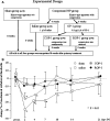
Effects of an intradiscal injection of OP-1 on the hyperalgesia induced by an application of the degenerative nucleus pulposus on the nerve root (With permission, Kawakami M et al. Osteogenic protein-1 (osteogenic protein-1/bone morphogenetic protein-7) inhibits degeneration and pain-related behavior induced by chronically compressed nucleus pulposus in the rat. Spine 2005;30:1933–9). a To degenerate discs, the apparatus was used to chronically apply compression to the tail. Four weeks after surgery, some discs received OP-1 injection (0.2 μg/1 μl) and either continuous compression was applied (COP-1) or the compression was released (ROP-1). The sham group received the initial surgery and was kept without compression. The saline group received the compression treatment and at 4 weeks, the disc received a saline injection. b Rats in the saline group showed evidence of mechanical hyperalgesia in the ipsilateral hind paws from 2 days to 2 weeks after surgery. In rats in the COP-1 and ROP-1 groups, which were treated with intradiscal injection of OP-1, mechanical hyperalgesia was not observed
Fig. 6
Fig. 6
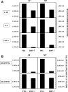
Intradiscal injections of recombinant human bone morphogenetic protein-7 (BMP-7/OP-1) significantly suppressed the mRNA expression of cytokines and catabolic enzymes in the rabbit anular puncture model (With permission, Pichika R et al. Intradiscal injection of recombinant human bone morphogenetic protein-7 significantly suppressed the expression of cytokines and catabolic enzymes in the rabbit anular puncture model. Ortho Res Soc Trans 33:1389, 2008). Four weeks after the anular puncture, the rabbits received an injection of phosphate buffered saline (10 μl, PBS group, n = 12 discs) or rhBMP-7 (100 μg, in 10 μl PBS, BMP-7 group) into the center of the NP. At the 12-week time point after the injection, AF and NP tissues were isolated and subjected to the quantitative PCR using the gene-specific primers for IL-1β, TNF-α, IL-6, ADAMTS4 and ADAMTS5. A single injection of rhBMP-7 induced a significantly decreased expression of cytokines (IL-1β, IL-6 and TNF-α, a) and ADAMTS4 mRNA (b)

Similar articles

Cited by

References

    1. Masuda K, Oegema TR, Jr, An HS. Growth factors and treatment of intervertebral disc degeneration. Spine. 2004;29:2757–2769. doi: 10.1097/01.brs.0000146048.14946.af. - DOI - PubMed
    1. Masuda K, An HS. Growth factors and the intervertebral disc. Spine J. 2004;4:330S–340S. doi: 10.1016/j.spinee.2004.07.028. - DOI - PubMed
    1. Ahn SH, Cho YW, Ahn MW, Jang SH, Sohn YK, Kim HS. mRNA expression of cytokines and chemokines in herniated lumbar intervertebral discs. Spine. 2002;27:911–917. doi: 10.1097/00007632-200205010-00005. - DOI - PubMed
    1. Burke JG, Watson RW, Conhyea D, McCormack D, Dowling FE, Walsh MG, Fitzpatrick JM. Human nucleus pulposus can respond to a pro-inflammatory stimulus. Spine. 2003;28:2685–2693. doi: 10.1097/01.BRS.0000103341.45133.F3. - DOI - PubMed
    1. Kang JD, Georgescu HI, McIntyre-Larkin L, Stefanovic-Racic M, Donaldson WF, 3rd, Evans CH. Herniated lumbar intervertebral discs spontaneously produce matrix metalloproteinases, nitric oxide, interleukin-6, and prostaglandin E2. Spine. 1996;21:271–277. doi: 10.1097/00007632-199602010-00003. - DOI - PubMed

MeSH terms

Substances