

Asymmetry and dyslexia
- PMID: 19005910
- PMCID: PMC2586924
- DOI: 10.1080/87565640802418597
Asymmetry and dyslexia
Abstract
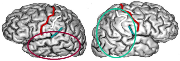
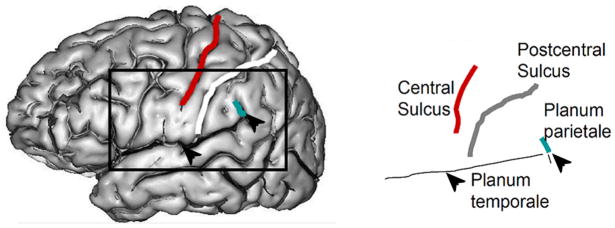
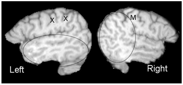
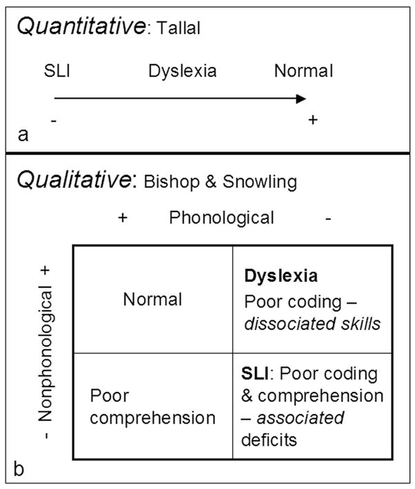
Developmental language disorders are characterized by a maturational trajectory that deviates or lags that of normal children. Given the wide variation in the rate of normal language development, diagnosis and classification of these disorders poses severe problems for the clinician. Our laboratory has been searching for anatomical signatures that could aid the development of a neurobiologically based classification. Quantitative analysis of the magnetic resonance imaging (MRI) brain scans of a series of samples of children and adults with reading and language disorders has identified two clusters with contrasting anatomical and reading profiles. Individuals with small symmetrical brain structures tend to have deficits in multiple domains of written and oral language whereas those with larger asymmetrical structures are more likely to have the isolated phonological deficits seen in adults with compensated dyslexia. Surprisingly, the anatomical risk factors that define these clusters do not form a continuum of increasing severity but deviate in opposite directions from normal. Individuals with moderate brain size and asymmetry typically demonstrate the best overall performance. Further research should determine if phonological impairments in the two clusters are associated with differing genetic and environmental risk factors requiring different types of intervention.
Figures
Similar articles
-
Individual differences in anatomy predict reading and oral language impairments in children.Brain. 2006 Dec;129(Pt 12):3329-42. doi: 10.1093/brain/awl262. Epub 2006 Sep 29. Brain. 2006. PMID: 17012292
-
White matter alterations and tract lateralization in children with dyslexia and isolated spelling deficits.Hum Brain Mapp. 2019 Feb 15;40(3):765-776. doi: 10.1002/hbm.24410. Epub 2018 Sep 29. Hum Brain Mapp. 2019. PMID: 30267634 Free PMC article.
-
Anatomical risk factors that distinguish dyslexia from SLI predict reading skill in normal children.J Commun Disord. 2002 Nov-Dec;35(6):501-31. doi: 10.1016/s0021-9924(02)00120-x. J Commun Disord. 2002. PMID: 12443050
-
Neuroanatomical markers for dyslexia: a review of dyslexia structural imaging studies.Neuroscientist. 2004 Aug;10(4):362-71. doi: 10.1177/1073858404263596. Neuroscientist. 2004. PMID: 15271263 Review.
-
Dyslexia (specific reading disability).Biol Psychiatry. 2005 Jun 1;57(11):1301-9. doi: 10.1016/j.biopsych.2005.01.043. Biol Psychiatry. 2005. PMID: 15950002 Review.
Cited by
-
Atypical brain torque in boys with developmental stuttering.Dev Neuropsychol. 2012;37(5):434-52. doi: 10.1080/87565641.2012.661816. Dev Neuropsychol. 2012. PMID: 22799762 Free PMC article.
-
It's never too early to get it Right: A conserved role for the cytoskeleton in left-right asymmetry.Commun Integr Biol. 2013 Nov 1;6(6):e27155. doi: 10.4161/cib.27155. Epub 2013 Nov 14. Commun Integr Biol. 2013. PMID: 24505508 Free PMC article.
-
Functional Hemispheric (A)symmetries in the Aged Brain-Relevance for Working Memory.Front Aging Neurosci. 2018 Mar 12;10:58. doi: 10.3389/fnagi.2018.00058. eCollection 2018. Front Aging Neurosci. 2018. PMID: 29593523 Free PMC article.
-
A unified model for left-right asymmetry? Comparison and synthesis of molecular models of embryonic laterality.Dev Biol. 2013 Jul 1;379(1):1-15. doi: 10.1016/j.ydbio.2013.03.021. Epub 2013 Apr 10. Dev Biol. 2013. PMID: 23583583 Free PMC article. Review.
-
Individual differences in reading skill and language lateralisation: a cluster analysis.Laterality. 2012;17(2):225-51. doi: 10.1080/1357650X.2011.561860. Epub 2011 Jul 19. Laterality. 2012. PMID: 22385144 Free PMC article.
References
-
- Belin P, Zilbovicius M, Fontaine A, Crozier S, Thivard L. Lateralization of speech and auditory temporal processing. Journal of Cognitive Neuroscience. 1998;10:536–540. - PubMed
-
- Best M, Demb J. Normal planum temporale asymmetry in dyslexics with a magnocellular deficit. Neuroreport. 1999;10:607–612. - PubMed
-
- Binder JR, Frost JA, Hammeke TA, Bellgowan PS, Rao SM, Cox RW. Conceptual processing during the conscious resting state. A functional MRI study. J Cogn Neurosci. 1999;11:80–95. - PubMed
-
- Bishop DV, Snowling MJ. Developmental dyslexia and specific language impairment: same or different? Psychol Bull. 2004;130:858–886. - PubMed
-
- Bruck M. Persistence of dyslexics’ phonological awareness deficits. Developmental Psychology. 1992;28:874–886.
Publication types
MeSH terms
Grants and funding
LinkOut - more resources
Full Text Sources