

Galanin inhibition of voltage-dependent Ca(2+) influx in rat cultured myenteric neurons is mediated by galanin receptor 1
- PMID: 19006083
- PMCID: PMC2935699
- DOI: 10.1002/jnr.21923
Galanin inhibition of voltage-dependent Ca(2+) influx in rat cultured myenteric neurons is mediated by galanin receptor 1
Abstract
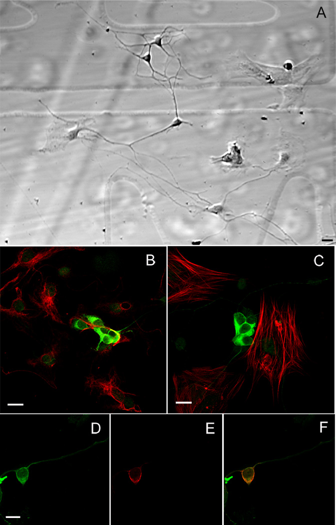
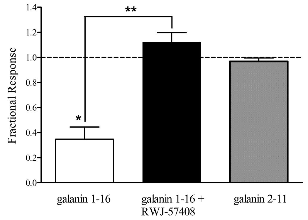
Galanin activates three receptors, the galanin receptor 1 (GalR1), GalR2, and GalR3. In the gastrointestinal tract, GalR1 mediates the galanin inhibition of cholinergic transmission to the longitudinal muscle and reduction of peristalsis efficiency in the small intestine. Galanin has also been shown to inhibit depolarization-evoked Ca2+ increases in cultured myenteric neurons. Because GalR1 immunoreactivity is localized to cholinergic myenteric neurons, we hypothesized that this inhibitory action of galanin on myenteric neurons is mediated by GalR1. We investigated the effect of galanin 1-16, which has high affinity for GalR1 and GalR2, in the presence or absence of the selective GalR1 antagonist, RWJ-57408, and of galanin 2-11, which has high affinity for GalR2 and GalR3, on Ca2+ influx through voltage-dependent Ca2+ channels in cultured myenteric neurons. Myenteric neurons were loaded with fluo-4 and depolarized by high K+ concentration to activate voltage-dependent Ca2+ channels. Intracellular Ca2+ levels were quantified with confocal microscopy. Galanin 1-16 (0.01-1 microM) inhibited the depolarization-evoked Ca2+ increase in a dose-dependent manner with an EC(50) of 0.172 microM. The selective GalR1 antagonist, RWJ-57408 (10 microM), blocked the galanin 1-16 (1 microM)-mediated inhibition of voltage-dependent Ca2+ channel. By contrast, the GalR2/GalR3 agonist, galanin 2-11 did not affect the K+-evoked Ca2+ influx in myenteric neurons. GalR1 immunoreactivity was localized solely to myenteric neurons in culture, as previously observed in intact tissue. These findings indicate that the inhibition of depolarization-evoked Ca2+ influx in myenteric neurons in culture is mediated by GalR1 and confirm the presence of functional GalR1 in the myenteric plexus. This is consonant with the hypothesis that GalR1 mediates galanin inhibition of transmitter release from myenteric neurons.
Figures


Similar articles
-
Galanin receptors in the rat gastrointestinal tract.Neuropeptides. 2005 Jun;39(3):349-52. doi: 10.1016/j.npep.2004.12.023. Neuropeptides. 2005. PMID: 16044511 Free PMC article. Review.
-
Galanin Receptors (GALR1, GALR2, and GALR3) Immunoexpression in Enteric Plexuses of Colorectal Cancer Patients: Correlation with the Clinico-Pathological Parameters.Biomolecules. 2022 Nov 27;12(12):1769. doi: 10.3390/biom12121769. Biomolecules. 2022. PMID: 36551197 Free PMC article.
-
Selective stimulation of GalR1 and GalR2 in rat substantia gelatinosa reveals a cellular basis for the anti- and pro-nociceptive actions of galanin.Pain. 2008 Jul;137(1):138-146. doi: 10.1016/j.pain.2007.08.030. Epub 2007 Oct 1. Pain. 2008. PMID: 17910903
-
Preferential activation by galanin 1-15 fragment of the GalR1 protomer of a GalR1-GalR2 heteroreceptor complex.Biochem Biophys Res Commun. 2014 Sep 26;452(3):347-53. doi: 10.1016/j.bbrc.2014.08.061. Epub 2014 Aug 22. Biochem Biophys Res Commun. 2014. PMID: 25152404
-
Regulation of limbic status epilepticus by hippocampal galanin type 1 and type 2 receptors.Neuropeptides. 2005 Jun;39(3):277-80. doi: 10.1016/j.npep.2004.12.003. Epub 2005 Jan 28. Neuropeptides. 2005. PMID: 15944022 Review.
Cited by
-
Review: Occurrence and Distribution of Galanin in the Physiological and Inflammatory States in the Mammalian Gastrointestinal Tract.Front Immunol. 2021 Jan 22;11:602070. doi: 10.3389/fimmu.2020.602070. eCollection 2020. Front Immunol. 2021. PMID: 33552060 Free PMC article. Review.
-
Glyphosate-induced changes in the expression of galanin and GALR1, GALR2 and GALR3 receptors in the porcine small intestine wall.Sci Rep. 2024 Apr 17;14(1):8905. doi: 10.1038/s41598-024-59581-8. Sci Rep. 2024. PMID: 38632282 Free PMC article.
-
Opioid-induced mitogen-activated protein kinase signaling in rat enteric neurons following chronic morphine treatment.PLoS One. 2014 Oct 10;9(10):e110230. doi: 10.1371/journal.pone.0110230. eCollection 2014. PLoS One. 2014. PMID: 25302800 Free PMC article.
-
Galanin inhibits GLP-1 and GIP secretion via the GAL1 receptor in enteroendocrine L and K cells.Br J Pharmacol. 2016 Mar;173(5):888-98. doi: 10.1111/bph.13407. Epub 2016 Feb 8. Br J Pharmacol. 2016. PMID: 26661062 Free PMC article.
-
Leptin modulates nutrient reward via inhibitory galanin action on orexin neurons.Mol Metab. 2015 Jul 16;4(10):706-17. doi: 10.1016/j.molmet.2015.07.002. eCollection 2015 Oct. Mol Metab. 2015. PMID: 26500842 Free PMC article.
References
-
- Anselmi L, Cervio E, Guerrini S, Vicini R, Agazzi A, Dellabianca A, Reeve JR, Jr, Tonini M, Sternini C. Identification of galanin receptor 1 on excitatory motor neurons in the guinea pig ileum. Neurogastroenterol Motil. 2005a;17(2):273–280. - PubMed
-
- Bauer FE, Zintel A, Kenny MJ, Calder D, Ghatei MA, Bloom SR. Inhibitory effect of galanin on postprandial gastrointestinal motility and gut hormone release in humans. Gastroenterology. 1989;97(2):260–264. - PubMed
Publication types
MeSH terms
Substances
Grants and funding
LinkOut - more resources
Full Text Sources
Molecular Biology Databases
Miscellaneous
