. 2009 Jan 20;512(3):305-22.
doi: 10.1002/cne.21903.

Secondary motoneurons in juvenile and adult zebrafish: axonal pathfinding errors caused by embryonic nicotine exposure

Affiliations
Free PMC article

Secondary motoneurons in juvenile and adult zebrafish: axonal pathfinding errors caused by embryonic nicotine exposure

Evdokia Menelaou et al. J Comp Neurol. .
Free PMC article

Abstract

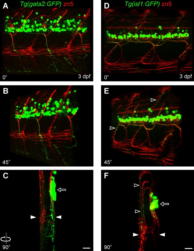
Nicotine is a drug of abuse that has been reported to have many adverse effects on the developing nervous system. We previously demonstrated that embryonic exposure to nicotine alters axonal pathfinding of spinal secondary motoneurons in zebrafish. We hypothesize that these changes will persist into adulthood. The Tg(isl1:GFP) line of zebrafish, which expresses green fluorescent protein (GFP) in a subtype of spinal secondary motoneurons, was used to investigate potential long-term consequences of nicotine exposure on motoneuron development. Anatomical characterization of Tg(isl1:GFP) zebrafish ranging between 3 and 30 days postfertilization (dpf) was initially performed in fixed tissue to characterize axonal trajectories in larval and juvenile fish. Tg(isl1:GFP) embryos were transiently exposed to 5-30 microM nicotine. They were then rescued from nicotine and raised into later stages of life (3-30 dpf) and fixed for microscopic examination. Morphological analysis revealed that nicotine-induced abnormalities in secondary motoneuron anatomy were still evident in juvenile fish. Live imaging of Tg(isl1:GFP) zebrafish using fluorescent stereomicroscopy revealed that the nicotine-induced changes in motoneuron axonal pathfinding persisted into adulthood. We detected abnormalities in 37-dpf fish that were transiently exposed to nicotine as embryos. These fish were subsequently imaged over a 7-week period of time until they were approximately 3 months of age. These pathfinding errors of spinal secondary motoneuron axons detected at 37 dpf persisted within the same fish until 86 dpf, the latest age analyzed. These findings indicate that exposure to nicotine during embryonic development can have permanent consequences for motoneuron anatomy in zebrafish.


Figures

Figure 1
Figure 1
Illustration depicting imaging methodology in fixed tissue. Zebrafish at various ages were analyzed via fluorescent microscopy with the aid of an ApoTome. All zebrafish were mounted on the side. Image stacks were acquired within the region denoted by the dashed box (top) and subsequent examinations using Imaris software included volume rendering allowing rotations in any direction. All rotations shown in this work are presented as horizontal rotations of the dorsoventral axis (middle, black arrow indicates rotational direction) with rostral regions moving away (bottom, left open arrow) and caudal regions moving out of the plane of the page (bottom, right open arrow).
Figure 2
Figure 2
Zn5 labeling of secondary motoneuron somata and their axons in isl1 and gata2 zebrafish. A: Photomicrograph of a projected z-stack from a 72 hpf isl1 zebrafish reveals GFP-positive motoneuron somata located in ventral spinal cord. Secondary motoneurons extend their axons dorsally to innervate dorsal musculature (filled arrow). B: Zn5-positive axons extend both ventrally and dorsally into the myotome. C: The merged image reveals that dorsally projecting GFP-positive axons are recognized by zn5. The GFP signal in spinal cord was saturated to reveal motoneuron axons. D: Projected z-stack from a 72-hpf gata2 zebrafish reveals GFP-positive motoneuron somata and ventrally projecting axons (filled arrowhead) and zn5-positive axons extending both ventrally and dorsally into the myotome. The GFP signal in spinal cord was saturated to reveal motoneuron axons. E,F: Expanded views from D and C merged images, respectively, show zn5 labeling of ventrally projecting GFP-positive motoneuron axons in a gata2 larva (left) and on dorsally projecting GFP-positive motoneuron axons in an isl1 larva (right). GFP-positive cells located more dorsally are presumably interneurons (open arrowhead). GI: Single focal plane images selected from a series of stacks from isl1, gata2, and isl1/gata2 zebrafish indicating that zn5 labels a population of motoneurons other than just the GFP-positive motoneurons in isl1 or gata2 zebrafish. Motoneuron somata that express GFP driven by the islet1 promoter (G) are detected by zn5 and are positioned in mid-dorsal spinal cord. Only a subpopulation of zn5-positive motoneuron somata, which are located in ventral spinal cord, is GFP-positive in gata2 zebrafish (H). Some interneurons located dorsally are not zn5-positive (open arrowheads). Zn5 immunoreactivity in isl1/gata2 transgenic zebrafish (I) detects most GFP-positive motoneurons. The presence of zn5-positive/GFP-negative cells indicates the presence of other secondary motoneuron subpopulations. A total of 21 fish were analyzed for this figure. A magenta/green copy of this figure is available as Supplemental Figure 3. Scale bars = 20 μm. A–C share the scale bar in C and G–I share the scale bar in I.
Figure 3
Figure 3
Secondary motoneuron axons in isl1 and gata2 zebrafish. A: Lateral view of a maximum projection from a z-stack (0° rotation) from a 72-hpf gata2 zebrafish labeled with zn5 reveals ventrally projecting motoneuron nerves exhibiting secondary and tertiary branches. B: Three-dimensional rendering at a 45° rotation reveals axons exiting ventral spinal cord that exhibit branching extending laterally within the myotome. C: At a 90° rotation the spinal cord (half spinal hemisegment) is to the right (open arrow) with the most superficial (lateral, zn5-positive) side of the fish to the left. Note the extensive branching from the ventral main nerve within the myotome (GFP signal below the white arrowheads). D: Lateral view (0° rotation) from a z-stack from a 72-hpf isl1 zebrafish reveals zn5 immunoreactivity in dorsally and ventrally projecting axons in addition to the slow muscle boundaries. E: Three-dimensional rendering at a 45° rotation reveals zn5-positive axons that project to reach the lateral edge and then take a dorsal path along the segmental boundaries at the most lateral part of the fish (open arrowheads). F: The 90° rotation reveals the cross-sectional view showing the left side of the spinal cord to the right (open arrow) with the superficial/lateral side of the fish to the left (open arrowheads). Both zn5- and GFP-positive axons are evidently extending from a medial plane (ventral of spinal cord) out into the most lateral surface of the fish (open arrowheads in E,F). Note the GFP-positive axonal trajectories located within the dorsal myotome (region above the white arrowheads). A total of 21 fish were analyzed for this figure. White arrowheads indicate the midline in C and F. A magenta/green copy of this figure is available as Supplemental Figure 4. Scale bars = 20 μm. A–C share the scale bar in C and D–F share the scale bar in F.
Figure 4
Figure 4
Axonal trajectories in isl1 zebrafish are associated with laterally located muscle fibers. A: Photomicrograph of 72-hpf wildtype larval zebrafish labeled with the antibody F59 to detect slow muscle fibers located at the most superficial part of the fish under the skin. B: Zn5 immunoreactivity localizes on superficial muscle cells only on their surfaces apposed to other slow muscle cells. C: The merged image reveals double antibody staining with F59/zn5. D: In 72-hpf isl1 zebrafish, GFP-positive secondary motoneuron axons extend into the dorsolateral myotome as shown by localization of the zn5 signals with GFP-positive axons. E: A magnified view of the boxed area shown in D demonstrates close association of the lateral most GFP-positive axons with zn5 signals (white arrows). F: Rh-α-btx labeling reveals muscle nicotinic acetylcholine receptors at the lateral region of the fish along zn5-positive segmental boundaries. G: Magnified lateral view of the boxed area shown in F shows the distribution of muscle nicotinic acetylcholine receptors at the lateral myotome. H: Side view (90° rotation) of the area shown in G reveals the spatial distribution of the muscle nicotinic acetylcholine receptors both at the slow (filled arrowheads) and fast muscle (open arrowheads) levels. Superficial/lateral side is to the left and medial is to the right. A total of 16 fish were analyzed for this figure. A magenta/green copy of this figure is available as Supplemental Figure 5. Scale bars = 25 μm in A–C; 20 μm in D–F; 5 μm in G. A–C share the scale bar in C.
Figure 5
Figure 5
Anatomical characterization of secondary motoneuron axons in isl1 zebrafish. The developmental progression of secondary motoneuron axons was examined and four distinct secondary motoneuron axon subpopulations were classified based on their anatomical position. AF: Projected images from a 3-dpf (A–C) and a 24-dpf isl1 zebrafish (D–F) are shown at 27° (A,D), 54° (B,E), and 90° (C,F) rotations. One population of axons exits ventral spinal cord from mid-segmental roots and then takes a dorsal turn (“check”-like) (open arrowheads) to extend and innervate musculature within dorsomedial myotome (filled arrowheads). A second population of axons (“loop”-like trajectory) projects ventrally to the midline, then it takes a lateral turn to reach the most lateral periphery and finally project dorsolaterally. A third class of axons exits from ventral spinal cord and continues to project ventromedially passing the midline to innervate the ventral-most musculature of the fish (filled arrows, D,F). At later stages in development, a fourth spatially distinct axonal trajectory is present, extending ventrally at the most lateral part of the fish (open arrows, E,F). G: A projected image (49° rotation) of a 24-dpf zebrafish reveals that the axons extending dorsoventrally at the most lateral region of the fish are originating from the same nerve (“loop”-like). H: Magnified view of the boxed area in G reveals the divergence of the two axonal trajectories (dashed arrows), each taking their own path innervating either dorsolateral or ventrolateral musculature. Scale bars = 20 μm. A–C share the scale bar in C and D–F share the scale bar in F.
Figure 6
Figure 6
Diagrammatic illustration of the motoneuron axons in isl1 zebrafish. Cartoons depict the patterning of the secondary motoneuron axons in the isl1 zebrafish according to our characterization shown in Figure 5. GFP-positive motoneuron somata located in ventral spinal cord are shown in green. Dotted line indicates the horizontal myoseptum. Continuous black lines (V-shaped) represent segmental boundaries. At 3 dpf, a subset of motoneuron somata extend their axons to exit from mid-segmental roots, where they travel shortly in a ventral path and then take a dorsomedial turn (red). Another population of secondary motoneurons axons (shown in blue) exits the same mid-segmental root to follow a ventral path until they reach the horizontal myoseptum. There they turn laterally and finally take a dorsolateral path. At 24 dpf, the motoneuron axons shown in red and blue at 3 dpf maintain their trajectories with the exception that the nerve bundles extending dorsomedially (red) at 24 dpf are putting out secondary and tertiary branches in the dorsomedial myotome. At later stages of development, another nerve is located ventrally and retains a ventromedial trajectory (orange). The motoneuron axons, shown in dashed blue, are diverging from the motoneuron axons shown in blue (dorsolateral) and they take a ventrolateral path. A total of 45 fish between 3–30 dpf were analyzed in generating this model. Cartoons are not drawn to scale. A magenta/green copy of this figure is available as Supplemental Figure 6.
Figure 7
Figure 7
Embryonic nicotine exposure causes secondary motoneuron axonal pathfinding errors early in development. A: Projected image from a z-stack shows a stage-matched control isl1 larval zebrafish (n = 15 fish) that has the characteristic pattern of the “check”-like trajectories and the ventrally projecting axons that loop laterally to take a dorsolateral path in each segment. B: Isl1 zebrafish embryos exposed to 5 μM nicotine (n = 5 fish) have abnormal trajectories exhibiting duplicated dorsal axons (open arrowhead) but no distinct abnormalities in the main axonal trajectories are observed. C: 15 μM nicotine exposure (n = 7 fish) causes pathfinding errors when axons reach the lateral periphery of the fish (open arrow). The axons do not properly loop dorsally and they exhibit extensive branching. D,E: 30 μM nicotine exposure (n = 6 fish) produces various axonal pathfinding errors including failure to project dorsal axons in some segments (D, filled arrow). Pathfinding abnormalities associated with axons extending out along the dorsolateral path and abnormal branching at the ventral root (E, open arrowheads) are also evident. Scale bars = 20 μm.
Figure 8
Figure 8
Nicotine-induced abnormalities revealed 3 weeks postexposure in fixed tissue. Isl1 zebrafish embryos exposed to nicotine were raised to 24 dpf for image examination. A: Stage-matched controls (n = 4 fish), exhibit the characteristic trajectories with the “loop”-like (arrowheads) and the “check”-like patterns (open arrow). B,C: Nicotine exposure (15–30 μM; n = 5 fish) results in abnormal motoneuron trajectories with extra axons exiting spinal cord in between segments (open arrowheads in B). In some cases, duplicated “check”-like trajectories were present (white arrow in C). Also, abnormalities were present at the most lateral region of the fish where the “loop”-like nerve diverges into two distinct axonal trajectories to extend either dorsolaterally or ventrolaterally (circle in C). This disorganization at the most lateral periphery was one of the most frequently encountered nicotine-induced phenotypes. Scale bars = 40 μm.
Figure 9
Figure 9
Nicotine-induced abnormalities at the lateral region of juvenile zebrafish. Isl1 zebrafish embryos exposed to nicotine were raised to 24 dpf and 30 dpf. Images shown are from a series of stacked images focusing on the most lateral motoneuronal trajectories. AC: Images of the most lateral motoneuron axonal trajectories in 24-dpf control (A,B, n = 4 fish) and nicotine-exposed (C, n = 5 fish) isl1 zebrafish. Arrowheads in A and B indicate the distinct choice point at the midline where these axonal trajectories diverge to follow their appropriate paths, dorsally and ventrally, along the segmental boundaries. Embryonic exposure to 30 μM nicotine causes axonal abnormalities at this lateral most region of 24-dpf isl1 zebrafish. The distinct choice point at the midline is not evident or clear in nicotine-exposed zebrafish where no center point can be distinguished. Rather a highly disorganized pattern is observed which is denoted by the white circle. D: Image of a 30-dpf unexposed control isl1 (n = 4 fish) showing the lateral axonal trajectories with a distinct choice point at the midline indicated by arrowheads. E: 30 dpf nicotine-exposed isl1 zebrafish (n = 6 fish) exhibit severe disorganization at this most lateral region of the fish as seen at 24 dpf. This distinct point is not evident in nicotine-exposed zebrafish and is denoted by the white circle to emphasize this abnormal phenotype observed. Scale bars = 40 μm.
Figure 10
Figure 10
In vivo live imaging in isl1 zebrafish at 17 dpf reveals nicotine-induced secondary motoneuron axon pathfinding errors. A: Representative image of an unexposed control (n = 5) shows the distinct motoneuron axons that project ventrally and then turn laterally (“loop”-like) (open arrowheads) to project into the dorsal musculature. The open arrows point to the “check”-like trajectories that project dorsally and innervate medially located musculature in every segment. B,C: Photomicrographs of isl1 zebrafish exposed to 15 μM nicotine (n = 5) show a duplicated axonal trajectory (B, filled arrowheads) and extra axons that exit ventral spinal cord in between segments (C, filled arrowhead). D,E: Isl1 zebrafish exposed to 30 μM nicotine (n = 6) exhibit varying abnormal trajectory phenotypes including extra branching medially between segments (D, filled arrow) and stalling of axons extending into the periphery (E, filled arrow). Scale bars = 40 μm.
Figure 11
Figure 11
The nicotine-induced abnormalities detected with in vivo live imaging are confirmed in fixed tissue. A: Photomicrograph of a 17-dpf isl1 zebrafish obtained during a live imaging session using fluorescent stereomicroscopy. A duplicated axonal trajectory appears to exit ventral spinal cord via an inter-segmental root and extends to the horizontal myoseptum where the “loop”-like axons take either a dorsolateral or ventrolateral path (filled arrowheads). The same fish was raised until 24 dpf and subsequently analyzed using fluorescent widefield microscopy to obtain z-stacks of images. B: Projected image at 24 dpf in the same segments corresponding to the ones observed during live imaging at 17 dpf (A) reveals abnormalities in axonal trajectories (filled arrowheads). A varicosity-like GFP signal (open arrowhead) was evident during live imaging in 17 dpf fish and most likely this GFP signal represents the turning point of an abnormal trajectory which appears to take a sharp dorsal turn within the myotome (open arrow). Scale bars = 40 μm.
Figure 12
Figure 12
Tracking embryonic nicotine-induced motoneuron axonal changes in adult zebrafish using live imaging in vivo. Isl1 zebrafish embryos embryonically exposed to 30 μM nicotine were raised into adulthood and monitored for anatomical changes during motoneuron development. Nicotine-exposed (n = 9 fish) and stage-matched controls (n = 5 fish) zebrafish were imaged weekly starting at 37 dpf for 7 weeks (86 dpf). AC: Representative photomicrographs of stage-matched controls at 37, 51, and 72 dpf, respectively. DF: Nicotine-exposed fish shown at 37, 51, and 72 dpf, respectively. G,H: Cartoon depicts normal patterns of axonal trajectories in unexposed zebrafish and two examples of nicotine-induced abnormal phenotypes. During these later developmental stages, the unexposed controls have a distinct pattern of dorsally and ventrally projecting axons located at the most lateral part of the fish. This region (black circle in A) corresponds to the region denoted by white arrowheads shown in Figure 9A,B. A varicosity-like GFP signal (GFP “center spot”) (A–C, filled arrows) is evident with additional axons branching from the “center spot” out between segments to where the “loop”-like trajectories extend laterally (perpendicular and out the plane of the page) from the medial myotome. In nicotine-exposed fish, these motoneuron axons have a disorganized patterning associated with the center point at the most lateral periphery. Axons also exhibit crossing over into adjacent segments (D–F, arrowheads) when compared to their stage-matched controls. Cartoon in D is not shown to scale. Scale bars = 100 μm.

Similar articles

Cited by

References

    1. Beattie CE, Hatta K, Halpern ME, Liu H, Eisen JS, Kimmel CB. Temporal separation in the specification of primary and secondary motoneurons in zebrafish. Dev Biol. 1997;187:171–182. - PubMed
    1. Berger F, Gage FH, Vijayaraghavan S. Nicotinic receptor-induced apoptotic cell death of hippocampal progenitor cells. J Neurosci. 1998;18:6871–6881. - PMC - PubMed
    1. Bhatt DH, Otto SJ, Depoister B, Fetcho JR. Cyclic AMP-induced repair of zebrafish spinal circuits. Science. 2004;305:254–258. - PubMed
    1. Burns FR, von Kannen S, Guy L, Raper JA, Kamholz J, Chang S. DM-GRASP, a novel immunoglobulin superfamily axonal surface protein that supports neurite extension. Neuron. 1991;7:209–220. - PubMed
    1. Chen Q, Sealock R, Peng HB. A protein homologous to the Torpedo postsynaptic 58K protein is present at the myotendinous junction. J Cell Biol. 1990;110:2061–2071. - PMC - PubMed

Publication types

MeSH terms

LinkOut - more resources