. 2009 Jan 20;512(3):399-418.
doi: 10.1002/cne.21896.

Axospinous synaptic subtype-specific differences in structure, size, ionotropic receptor expression, and connectivity in apical dendritic regions of rat hippocampal CA1 pyramidal neurons

Affiliations

Axospinous synaptic subtype-specific differences in structure, size, ionotropic receptor expression, and connectivity in apical dendritic regions of rat hippocampal CA1 pyramidal neurons

Daniel A Nicholson et al. J Comp Neurol. .

Abstract

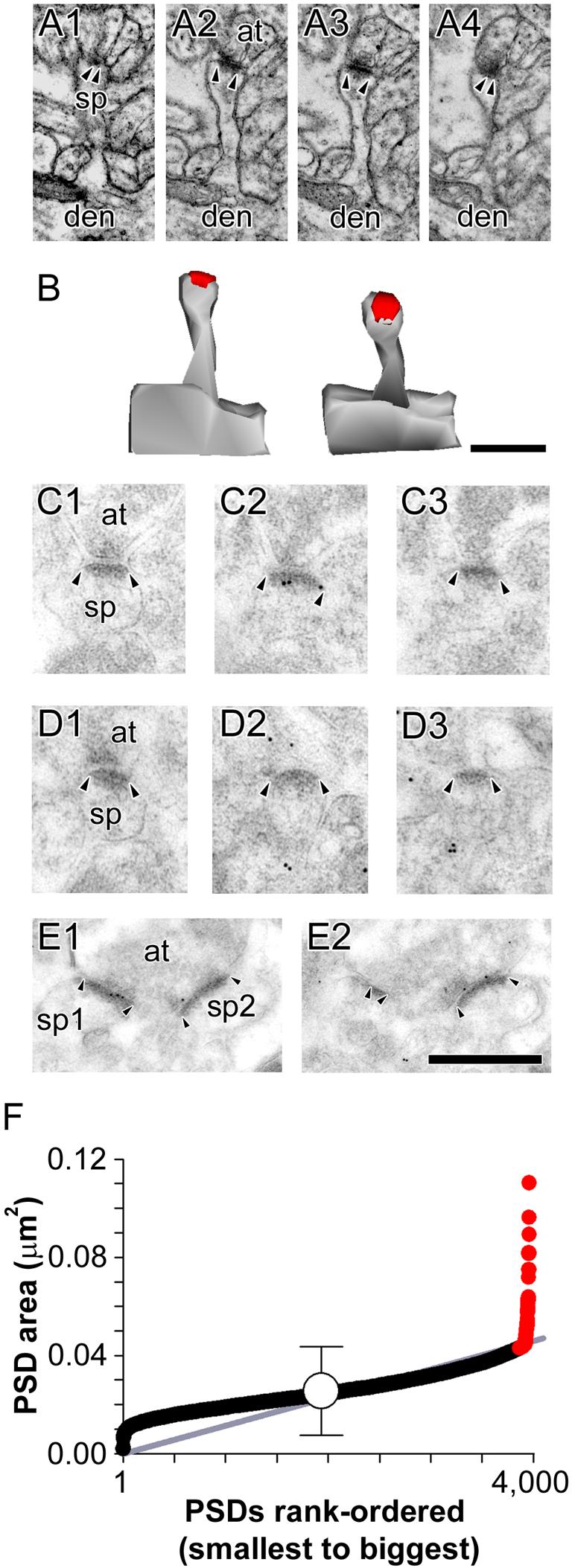
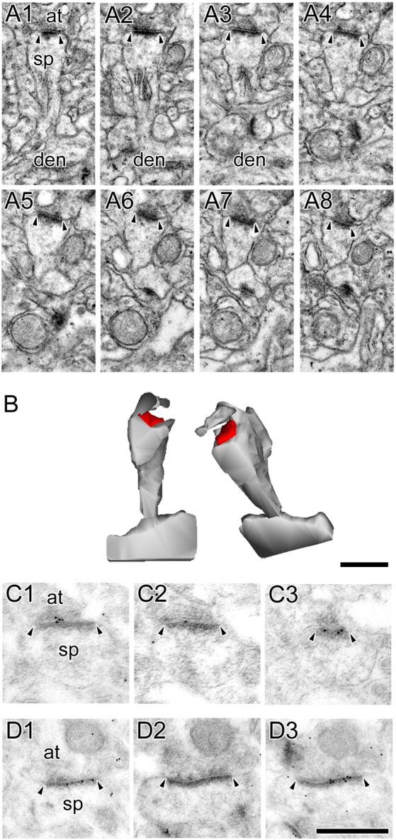
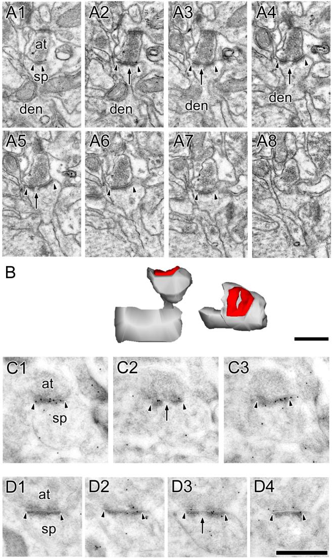
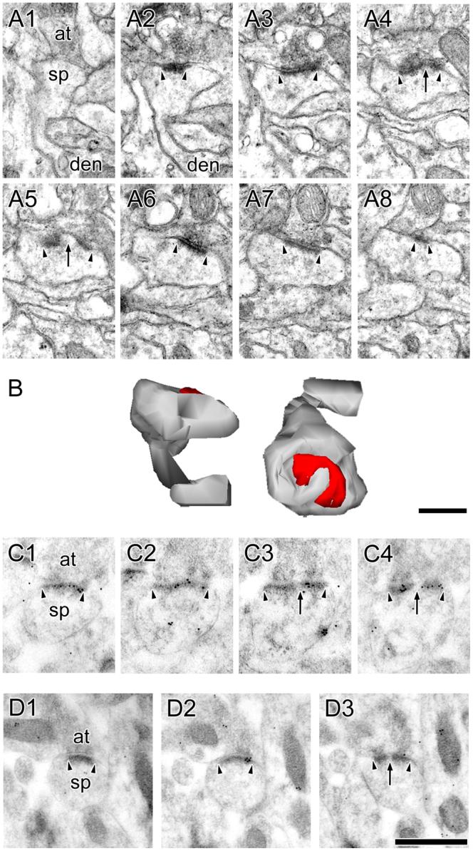
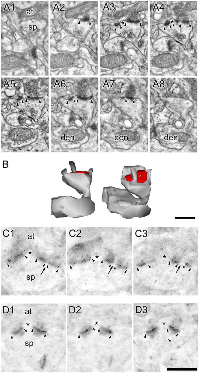
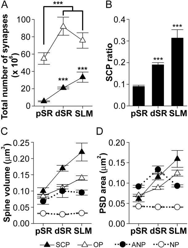
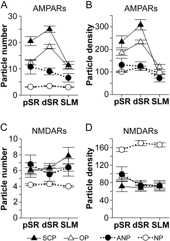
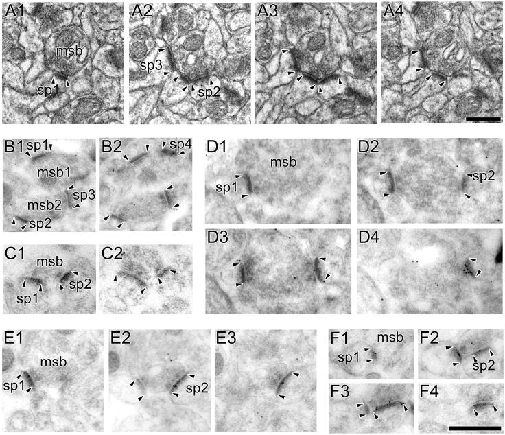
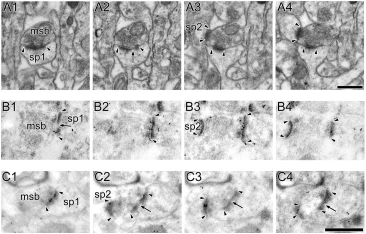
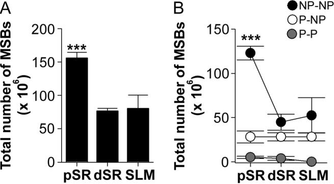
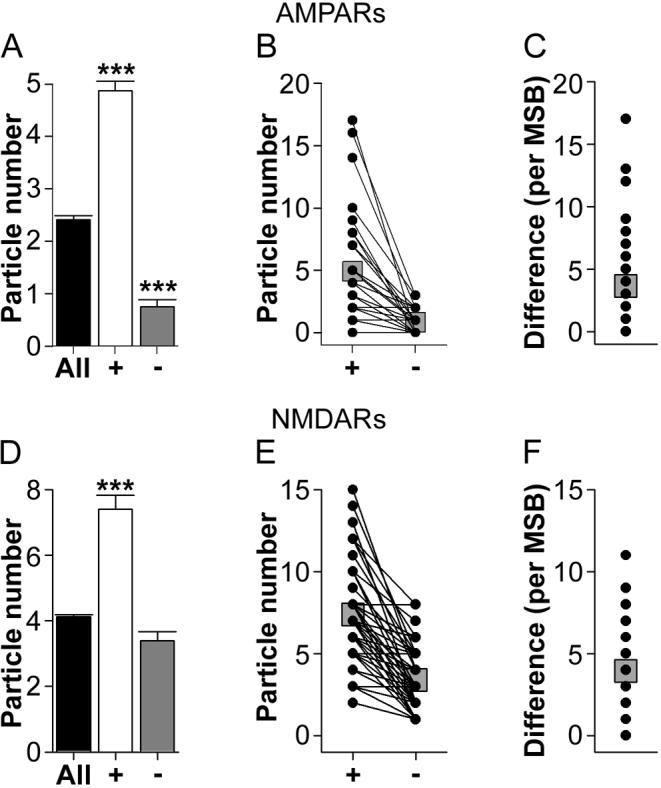
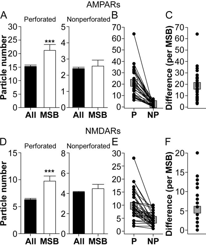
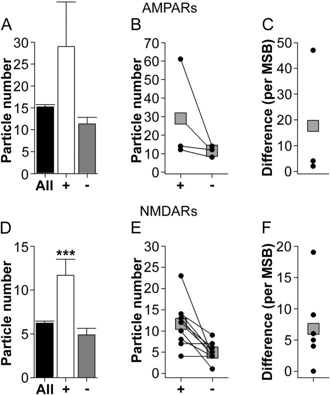
The morphology of axospinous synapses and their parent spines varies widely. Additionally, many of these synapses are contacted by multiple synapse boutons (MSBs) and show substantial variability in receptor expression. The two major axospinous synaptic subtypes are perforated and nonperforated, but there are several subcategories within these two classes. The present study used serial section electron microscopy to determine whether perforated and nonperforated synaptic subtypes differed with regard to their distribution, size, receptor expression, and connectivity to MSBs in three apical dendritic regions of rat hippocampal area CA1: the proximal and distal thirds of stratum radiatum, and the stratum lacunosum-moleculare. All synaptic subtypes were present throughout the apical dendritic regions, but there were several subclass-specific differences. First, segmented, completely partitioned synapses changed in number, proportion, and alpha-amino-3-hydroxy-5-methyl-4-isoxazolepropionate (AMPA) receptor expression with distance from the soma beyond that found within other perforated synaptic subtypes. Second, atypically large, nonperforated synapses showed N-methyl-D-aspartate (NMDA) receptor immunoreactivity identical to that of perforated synapses, levels of AMPA receptor expression intermediate to that of nonperforated and perforated synapses, and perforated synapse-like changes in structure with distance from the soma. Finally, MSB connectivity was highest in the proximal stratum radiatum, but only for those MSBs composed of nonperforated synapses. The immunogold data suggest that most MSBs would not generate simultaneous depolarizations in multiple neurons or spines, however, because the vast majority of MSBs are comprised of two synapses with abnormally low levels of receptor expression, or involve one synapse with a high level of receptor expression and another with only a low level.


Figures

Figure 1
Figure 1. Electron micrographs of “typical” nonperforated synapses
A: Serial sections through a nonperforated (NP) synapse between an axon terminal (at) and a spine (sp) that is seen to connect to its parent dendrite (den). B: 3-dimensional reconstructions of the NP synapse and its parent spine in their original orientation (Left) and rotated (Right) to illustrate the continuous shape of its postsynaptic density (PSD). C: Serial sections through a NP synapse (arrowheads) that was immunopositive for AMPA-type receptors (AMPARs). D: Serial sections through a NP synapse (arrowheads) that was immunonegative for AMPARs. E: Serial sections through two NP synapses (sp1 and sp2; arrowheads) making contact with the same presynaptic axon terminal (at), both of which were immunopositive for NMDA-type receptors. Scale bars = 0.5 μm. F: Scatterplot of the PSD area for all nonperforated synapses in the present study. PSD areas are plotted in rank-order according to their size. Gray line indicates the linear regression that nonperforated synapses follow as they increase in size. Mean PSD size (white circle) is plotted ± 2 standard deviations (S.D.) to illustrate that a subset of NP synapses deviates from the linear trajectory (gray line) at ∼ 2 S.D. Such synapses (red circles) were considered atypically large nonperforated (ANP) synapses in the present study.
Figure 2
Figure 2. Electron micrographs of atypically large nonperforated synapses
A: Serial sections through an atypically large nonperforated (ANP) synapse (arrowheads) between an axon terminal (at) and a spine (sp), which is seen connecting to its parent dendrite (den). A spine apparatus is observed in the spine head/neck region. B: 3-dimensional reconstructions of the ANP synapse and its parent spine in their original orientation (Left) and rotated (Right) to illustrate the continuous shape and large size of its postsynaptic density (PSD). Note the nonsynaptic spinule emanating from a perisynaptic region of the spine head. C, D: Serial sections through ANP synapses between axon terminals (at) and spines (sp), which were always immunopositive for AMPA-type receptors (C) and NMDA-type receptors (D). Scale bars = 0.5 μm. Though the ANP synapses showing AMPA-type and NMDA-type are different synapses, all ANP synapses were immunopositive for both. It is likely, then, that each of these synapses, had they been immunostained for both AMPA-type and NMDA-type receptors, would be immunopositive for both. Also note that all but 1 of the immunogold particles in panel C1 are considered synaptic according to our criteria (i.e., on or otherwise within 20 nm of the postsynaptic density, or in the synaptic cleft).
Figure 3
Figure 3. Electron micrographs of perforated synapses with fenestrated postsynaptic densities
A: Serial sections through a perforated synapse with a fenestrated postsynaptic density (PSD; FP synapse) between a spine (sp) protruding from a dendrite (den) and an axon terminal (at). FP synapses are characterized by a hole or fenestration (arrows) in their PSD profile (arrowheads). B: 3-dimensional reconstructions of the FP synapse and its parent spine in their original orientation (Left) and rotated (Right) to illustrate the discontinuity in its PSD. C, D: Serial sections through two FP synapses between axon terminals (at) and dendritic spines (sp) showing that FP synapses are highly immunoreactive for AMPA-type receptors (C) and NMDA-type receptors (D). Scale bars = 0.5 μm.
Figure 4
Figure 4. Electron micrographs of perforated synapses with horseshoe-shaped postsynaptic densities
A: Serial sections through a perforated synapse with a horseshoe-shaped postsynaptic density (PSD; HP synapse) between a spine (sp), shown connecting to its parent dendrite (den), and an axon terminal (at). HP synapses are characterized by the appearance of PSD profiles that are initially separated by spine cytoplasm (arrows), but then subsequently can be seen as a single PSD (arrowheads). B: 3-dimensional reconstructions of the HP synapse and its parent spine in their original orientation (Left) and rotated (Right) to illustrate the discontinuity in its PSD. C, D: Serial sections through two HP synapses between axon terminals (at) and dendritic spines (sp) showing that HP synapses are highly immunoreactive for AMPA-type receptors (C) and NMDA-type receptors (D). Scale bars = 0.5 μm.
Figure 5
Figure 5. Electron micrographs of perforated synapses with segmented, completely partitioned postsynaptic densities
A: Serial sections through a perforated synapse with a segmented, completed partitioned (SCP) postsynaptic density (PSD; arrowheads) between a spine (sp) on a dendrite (den) and an axon terminal (at). SCP synapses are characterized by multiple PSD profiles (arrowheads), each of which is separated from the other by a complete spine partition (asterisk) that invaginates that presynaptic axon terminal. In the synapse shown, one of the PSD plates is itself discontinuous (arrow). Note the presence of a spine apparatus in the spine head/neck region. B: 3-dimensional reconstructions of the SCP synapse and its parent spine in their original orientation (Left) and rotated (Right) to illustrate that it is composed of two distinct PSD plates separated by a complete spine partition. C: Serial sections through a SCP synapse between an axon terminal (at) and a dendritic spine (sp). The AMPA-type receptor immunoreactivity of SCP synapses is significantly higher than that of other synaptic subtypes in hippocampal region CA1. PSD profiles of the SCP synapse (arrowheads) are separated by a complete spine partition (asterisk) on the one hand, and by a cytoplasmic region of the spine head on the other (arrows). D: Serial sections through a SCP synapse between a dendritic spine (sp) and an axon terminal (at), showing the SCP synapses express NMDARs. Note that the PSD plates (arrowheads) are separated from each other by a complete spine partition (asterisk). Note also that each PSD plate of the SCP synapses expresses both AMPARs and NMDARs. Scale bars = 0.5 μm.
Figure 6
Figure 6. Total number and ratio of segmented, completely partitioned synapses and spine/synapse morphology as a function of dendritic location
A: Total number (± standard error of the mean; S.E.M.) of segmented, completely partitioned (SCP) synapses (black triangles) and other types of perforated (OP) synapses (white triangles) in three regions of the apical dendrite: proximal stratum radiatum (pSR), distal stratum radiatum (dSR), and stratum lacunosum-moleculare (SLM). There were significantly more OP synapses in dSR and SLM as compared to pSR (asterisks). The number of SCP synapses significantly increased with distance from the soma (asterisks). B: The ratio (± S.E.M.) of SCP synapses as a function of all perforated synapses increased progressively with distance from the soma (asterisks). C: Spine volume for SCP synapses was highest, whereas that for typical nonperforated (NP; white circles) synapses was lowest in all three dendritic regions. Spine volume for OP and SCP synapses increased with distance from the soma, whereas neither NP nor atypically large nonperforated (ANP; black circles) synapses changed in size as a function of dendritic location. Spine sizes for ANP and OP synapses did not differ significantly from each other. D: Postsynaptic density (PSD) area for synapses in pSR, dSR, and SLM. SCP and OP synapses increased in size with distance from the soma, but SCP synapses were bigger in dSR and SLM. ANP synapses were largest in pSR, the same size as SCP synapses in dSR, and intermediate to that of NP and perforated synapses in SLM. NP synapses were the only subtype whose size remained constant in all dendritic regions.
Figure 7
Figure 7. Expression levels of AMPA-type and NMDA-type receptors among the axospinous synaptic subtypes as a function of distance from the soma
A: Immunogold particle number (per synapse) for AMPA-type receptors (AMPARs) for segmented, completely partitioned perforated synapses (SCP; black triangles), other types of perforated synapses (OP; white triangles), as well as nonperforated (NP; white circles) and atypically large nonperforated (ANP; black circles) synapses in three regions of the apical dendrites: proximal stratum radiatum (pSR), distal stratum radiatum (dSR), and stratum lacunosum-moleculare (SLM). SCP synapses had the highest number of particles for AMPARs in pSR and dSR. OP and ANP synapses had the same number of immunogold particles for AMPARs in pSR, but OP synapses had more in dSR. AMPAR immunoexpression did not differ with distance from the soma among NP and ANP synapses, but it increased among SCP and OP synapses between pSR and dSR. In SLM, however, OP and SCP synapses did not differ with respect to their AMPAR immunoreactivity. In dSR and SLM, the level of expression for AMPARs among ANP synapses was intermediate to that of OP and NP synapses. B: Density (per μm2 of postsynaptic density area) of immunogold particles for AMPARs in pSR, dSR, and SLM. Immunogold particle density did not differ between NP and ANP synapses, but OP and SCP synapses had a significantly higher particle density than both nonperforated synaptic subtypes. SCP synapses had the highest particle density in pSR and dSR, but particle density for both OP and SCP synapses increased between pSR and dSR. There were no statistically significant differences among synapses in SLM, except that NP synapses had the lowest particle density. C: Immunogold particle number (per synapse) for NMDA-type receptors (NMDARs) in pSR, dSR, and SLM. NMDAR expression did not change with distance from the soma, but NP synapses always had fewer than the other synaptic subtypes. ANP, OP, and SCP synapses did not differ significantly from each other in any dendritic region. D: Density (per μm2 of postsynaptic density area) of immunogold particles for NMDARs was highest among NP synapses. NMDAR immunogold particle density did not change as a function of distance from the soma, nor did it differ among ANP, OP, and SCP synapses.
Figure 8
Figure 8. Electron micrographs of multiple synapse boutons involving only nonperforated synapses
A: Serial sections through a multiple synapse bouton (MSB) involving only nonperforated synapses (NP-NP MSB). The axon terminal (msb) synapses with the different spines (sp1, sp2, and sp3), all of which have nonperforated postsynaptic densities (PSD; arrowheads). B: Two MSBs (msb1 and msb2) between synapses showing weak AMPA-type receptor (AMPAR) immunoreactivity. One synapse (sp1) does not have any immunogold particles for AMPARs projected onto its PSD, and the others (sp2, sp3, sp4) have only one immunogold particle on their PSD profile. C: A NP-NP MSB involving two NP synapses (sp1 and sp2; arrowheads), both of which lack any AMPAR immunoreactivity. D: A NP-NP MSB between an axon terminal (msb) and two spines (sp1 and sp2). One synapse has 9 immunogold particles for AMPARs (sp2), whereas the other (sp1) lacks them. E: A NP-NP MSB between two spines (sp1 and sp2), both of which are immunoreactive for NMDA-type receptors (NMDARs). F: A NP-NP MSB between two spines (sp1 and sp2), which have 3 (sp1) and 8 (sp2) immunogold particles for NMDARs, respectively. Scale bars = 0.5 μm.
Figure 9
Figure 9. Electron micrographs of multiple synapse boutons involving nonperforated and perforated synapses
A: Serial sections through a multiple synapse bouton (MSB) involving a perforated (sp1) and a nonperforated synapse (sp2; P-NP MSB). The perforated synapse (sp1) is identified by a discontinuity (arrow) in its postsynaptic density (PSD; arrowheads), whereas the PSD of the nonperforated synapse is continuous in all sections (arrowheads). B: A P-NP MSB between a perforated (sp1) and a nonperforated (sp2) synapse. The perforated synapse (sp1) has 16 immunogold particles for AMPA-type receptors projected onto its PSD, whereas the nonperforated synapse (sp2) has none. C: A P-NP MSB between a perforated (sp1) and a nonperforated (sp2) synapse, both of which are immunoreactive for NMDA-type receptors (NMDARs). The perforated synapse (sp1) shows a discontinuity (arrow) in its PSD profiles (arrowheads), whereas the nonperforated synapse has continuous PSD profiles (arrowheads). The perforated and nonperforated synapses have 7 and 6 immunogold particles for NMDARs projected onto their PSDs, respectively. Scale bars = 0.5 μm.
Figure 10
Figure 10. Electron micrographs of multiple synapse boutons involving only perforated synapses
A: Serial sections through a multiple synapse bouton (msb) involving only perforated synapses (sp1, sp2, and sp3; P-P MSB). Each synapse (arrowheads) has at least one discontinuity in its postsynaptic density (PSD) profile (arrows). B: A P-P MSB involving two perforated synapses (sp1 and sp2) that are immunoreactive for AMPA-type receptors (AMPARs). One synapse (sp1) has 12 immunogold particles for AMPARs projected onto its PSD, and the other (sp2) has 14. C: A P-P MSB involving two perforated synapses (sp1 and sp2) that have 5 (sp1) and 4 (sp2) immunogold particles for NMDA-type receptors. Scale bars = 0.5 μm.
Figure 11
Figure 11. Total number of multiple synapse boutons
A: Proximal stratum radiatum (pSR) had more multiple synapse boutons (MSBs) than both distal stratum radiatum (dSR) and stratum lacunosum-moleculare (SLM; asterisks). B: pSR had more MSBs involving exclusively nonperforated synapses (NP-NP MSB; black circles) than dSR or SLM (asterisks). The total number of MSBs involving either perforated and nonperforated synapses (P-NP MSB; white circles) or exclusively perforated synapses (P-P MSB; gray circles) did not differ among the three apical dendritic regions.
Figure 12
Figure 12. Expression of AMPA-type and NMDA-type receptors in synapses of multiple synapse boutons that involve only nonperforated synapses
A: Average immunogold particle number for AMPA-type receptors (AMPARs) for all nonperforated (NP) synapses in the present study (black bar), the synapse with the higher number of immunogold particles in each nonperforated synapse-only multiple synapse bouton (NP-NP MSB; plus sign), and the synapse with the lower number of immunogold particles in each NP-NP MSB (minus sign). Asterisks indicate that NP-NP MSBs, on average, involved a nonperforated synapse with an abnormally high level of AMPAR expression and one with a significantly lower level of AMPAR expression compared to the overall population of NP synapses. B: Immunogold particles for AMPARs for each synapse involved in each NP-NP MSB, plotted according to which synapse had a higher (plus sign) or lower (minus sign) number. C: The difference in the immunogold particle number for AMPARs between the synapses of each NP-NP MSB. D: Average immunogold particle number for NMDA-type receptors (NMDARs) for all nonperforated (NP) synapses in the present study (black bar), the synapse with the higher number of immunogold particles in each NP-NP MSB (plus sign), and the synapse with the lower number of immunogold particles in each NP-NP MSB (minus sign). Asterisks indicate that NP-NP MSBs, on average, involved a nonperforated synapse with an abnormally high level of NMDAR expression and one with an expression level typical of the overall population. E: Immunogold particles for NMDARs for each synapse involved in each NP-NP MSB, plotted according to which synapse had a higher (plus sign) or lower (minus sign) number. F: The difference in the immunogold particle number for NMDARs between the synapses of each NP-NP MSB. Means are plotted in B, C, E, and F as gray squares.
Figure 13
Figure 13. Expression of AMPA-type and NMDA-type receptors in synapses of multiple synapse boutons that involve a mix of nonperforated and perforated synapses
A: Left panel Average immunogold particle number for AMPA-type receptors (AMPARs) for all perforated synapses in the present study (black bar), and for the perforated synapses of each perforatednonperforated synapse multiple synapse bouton (P-NP MSB; white bar). The asterisk indicates that perforated synapses involved in P-NP MSBs have a higher level of AMPAR expression than would be expected from the overall population of perforated synapses. Right panel Average immunogold particle number for AMPARs for all nonperforated (NP) synapses in the present study (black bar), and for the NP synapses of each P-NP MSB (white bar). B: Immunogold particles for AMPARs for each synapse involved in each P-NP MSB, plotted according to whether its PSD was perforated (P) or nonperforated (NP). C: The difference in the immunogold particle number for AMPARs between the synapses of each P-NP MSB. D: Left panel Average immunogold particle number for NMDA-type receptors (NMDARs) for all perforated synapses in the present study (black bar), and for the perforated synapses of each P-NP MSB (white bar). The asterisk indicates that perforated synapses involved in P-NP MSBs have a higher level of NMDAR expression than would be expected from the overall population of perforated synapses. Right panel Average immunogold particle number for NMDARs for all NP synapses in the present study (black bar), and for the NP synapses of each P-NP MSB (white bar). E: Immunogold particles for NMDARs for each synapse involved in each P-NP MSB, plotted according to whether its PSD was perforated (P) or nonperforated (NP). F: The difference in the immunogold particle number for NMDARs between the synapses of each P-NP MSB. Means are plotted in B, C, E, and Fas gray squares.
Figure 14
Figure 14. Expression of AMPA-type and NMDA-type receptors in synapses of multiple synapse boutons that involve only perforated synapses
A: Average immunogold particle number for AMPA-type receptors (AMPARs) for all perforated synapses in the present study (black bar), the synapse with the higher number of immunogold particles in each perforated synapse-only multiple synapse bouton (P-P MSB; plus sign), and the synapse with the lower number of immunogold particles in each P-P MSB (minus sign). B: Immunogold particles for AMPARs for each synapse involved in each P-P MSB, plotted according to which synapse had a higher (plus sign) or lower (minus sign) number. C: The difference in the immunogold particle number for AMPARs between the synapses of each P-P MSB. D: Average immunogold particle number for NMDA-type receptors (NMDARs) for all perforated synapses in the present study (black bar), the synapse with the higher number of immunogold particles in each P-P MSB (plus sign), and the synapse with the lower number of immunogold particles in each P-P MSB (minus sign). E: Immunogold particles for NMDARs for each synapse involved in each P-P MSB, plotted according to which synapse had a higher (plus sign) or lower (minus sign) number. F: The difference in the immunogold particle number for NMDARs between the synapses of each P-P MSB. Means are plotted in B, C, E, and F as gray squares.

Similar articles

Cited by

References

    1. Alvarez VA, Sabatini BL. Anatomical and physiological plasticity of dendritic spines. Annu Rev Neurosci. 2007;30:79–97. - PubMed
    1. Bloodgood BL, Sabatini BL. Neuronal activity regulates diffusion across the neck of dendritic spines. Science. 2005;310:866–869. - PubMed
    1. Bourne J, Harris KM. Do thin spines learn to be mushroom spines that remember? Curr Opin Neurobiol. 2007;17:381–386. - PubMed
    1. Bourne JN, Harris KM. Balancing structure and function at hippocampal dendritic spines. Annu Rev Neurosci. 2008;31:47–67. - PMC - PubMed
    1. Bredt DS, Nicoll RA. AMPA receptor trafficking at excitatory synapses. Neuron. 2003;40:361–379. - PubMed

Publication types

Substances