

Mutations in NR2E3 can cause dominant or recessive retinal degenerations in the same family
- PMID: 19006237
- PMCID: PMC3658139
- DOI: 10.1002/humu.20858
Mutations in NR2E3 can cause dominant or recessive retinal degenerations in the same family
Abstract
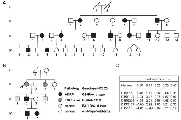
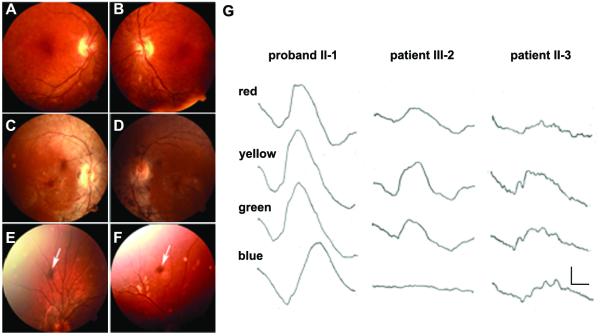
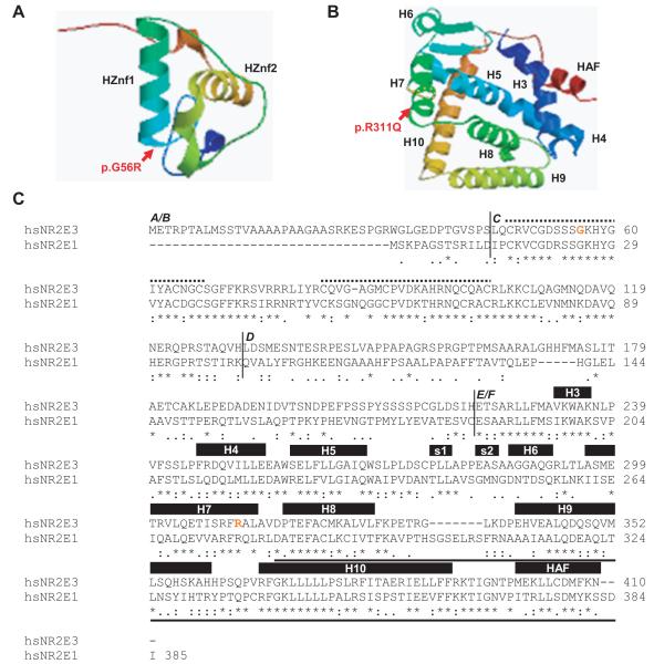
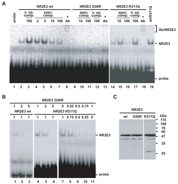
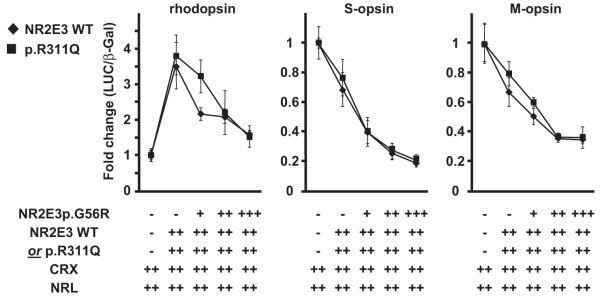
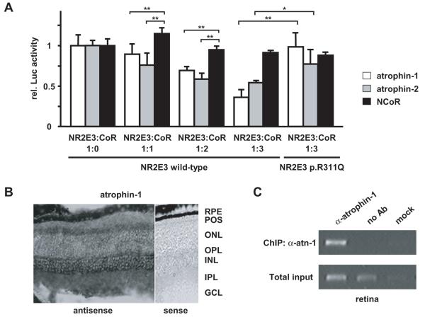
NR2E3, a photoreceptor-specific nuclear receptor (PNR), represses cone-specific genes and activates several rod-specific genes. In humans, mutations in NR2E3 have been associated with the recessively-inherited enhanced short-wavelength sensitive S-cone syndrome (ESCS) and, recently, with autosomal dominant (ad) retinitis pigmentosa (RP) (adRP). In the present work, we describe two additional families affected by adRP that carry a heterozygous c.166G>A (p.G56R) mutation in the NR2E3 gene. Functional analysis determined the dominant negative activity of the p.G56R mutant protein as the molecular mechanism of adRP. Interestingly, in one pedigree, the most common causal variant for ESCS (p.R311Q) cosegregated with the adRP-linked p.G56R mutation, and the compound heterozygotes exhibited an ESCS-like phenotype, which in 1 of the 2 cases was strikingly "milder" than the patients carrying the p.G56R mutation alone. Impaired repression of cone-specific genes by the corepressors atrophin-1 (dentatorubral-pallidoluysian atrophy [DRPLA] gene product) and atrophin-2 (arginine-glutamic acid dipeptide repeat [RERE] protein) appeared to be a molecular mechanism mediating the beneficial effect of the p.R311Q mutation. Finally, the functional dominance of the p.R311Q variant to the p.G56R mutation is discussed.
2008 Wiley-Liss, Inc.
Figures
Similar articles
-
Recurrent mutation in the first zinc finger of the orphan nuclear receptor NR2E3 causes autosomal dominant retinitis pigmentosa.Am J Hum Genet. 2007 Jul;81(1):147-57. doi: 10.1086/518426. Epub 2007 May 24. Am J Hum Genet. 2007. PMID: 17564971 Free PMC article.
-
Mutations in the DNA-binding domain of NR2E3 affect in vivo dimerization and interaction with CRX.PLoS One. 2009 Oct 12;4(10):e7379. doi: 10.1371/journal.pone.0007379. PLoS One. 2009. PMID: 19823680 Free PMC article.
-
Allele-Specific Knockout by CRISPR/Cas to Treat Autosomal Dominant Retinitis Pigmentosa Caused by the G56R Mutation in NR2E3.Int J Mol Sci. 2021 Mar 5;22(5):2607. doi: 10.3390/ijms22052607. Int J Mol Sci. 2021. PMID: 33807610 Free PMC article.
-
NR2E3 mutations in enhanced S-cone sensitivity syndrome (ESCS), Goldmann-Favre syndrome (GFS), clumped pigmentary retinal degeneration (CPRD), and retinitis pigmentosa (RP).Hum Mutat. 2009 Nov;30(11):1475-85. doi: 10.1002/humu.21096. Hum Mutat. 2009. PMID: 19718767 Review.
-
Clinical and Genetic Features of NR2E3-Associated Retinopathy: A Report of Eight Families with a Longitudinal Study and Literature Review.Genes (Basel). 2023 Jul 26;14(8):1525. doi: 10.3390/genes14081525. Genes (Basel). 2023. PMID: 37628579 Free PMC article.
Cited by
-
Loss of function mutations in RP1 are responsible for retinitis pigmentosa in consanguineous familial cases.Mol Vis. 2016 Jun 10;22:610-25. eCollection 2016. Mol Vis. 2016. PMID: 27307693 Free PMC article.
-
A comprehensive analysis of sequence variants and putative disease-causing mutations in photoreceptor-specific nuclear receptor NR2E3.Mol Vis. 2009 Oct 24;15:2174-84. Mol Vis. 2009. PMID: 19898638 Free PMC article.
-
Pathogenic mutations in TULP1 responsible for retinitis pigmentosa identified in consanguineous familial cases.Mol Vis. 2016 Jul 16;22:797-815. eCollection 2016. Mol Vis. 2016. PMID: 27440997 Free PMC article.
-
In pursuit of synthetic modulators for the orphan retina-specific nuclear receptor NR2E3.J Ocul Pharmacol Ther. 2013 Apr;29(3):298-309. doi: 10.1089/jop.2012.0135. Epub 2012 Oct 25. J Ocul Pharmacol Ther. 2013. PMID: 23098562 Free PMC article.
-
Dominant Retinitis Pigmentosa, p.Gly56Arg Mutation in NR2E3: Phenotype in a Large Cohort of 24 Cases.PLoS One. 2016 Feb 24;11(2):e0149473. doi: 10.1371/journal.pone.0149473. eCollection 2016. PLoS One. 2016. PMID: 26910043 Free PMC article.
References
-
- Akhmedov NB, Piriev NI, Chang B, Rapoport AL, Hawes NL, Nishina PM, Nusinowitz S, Heckenlively JR, Roderick TH, Kozak CA. A deletion in a photoreceptor-specific nuclear receptor mRNA causes retinal degeneration in the rd7 mouse. Proc. Natl. Acad. Sci. USA. 2000;97(10):5551–5556. others. - PMC - PubMed
-
- Audo I, Michaelides M, Robson AG, Hawlina M, Vaclavik V, Sandbach JM, Neveu MM, Hogg CR, Hunt DM, Moore AT. Phenotypic Variation in Enhanced S-cone Syndrome. Invest. Ophthalmol. Vis. Sci. 2008;49(5):2082–2093. others. - PubMed
-
- Bouayed-Tiab L, Delarive T, Agosti C, Borruat F-X, Munier FL, Schorderet DF. A Heterozygous Mutation in the NR2E3 Gene Is Associated With an Autosomal Dominant Retinitis Pigmentosa. Invest. Ophthalmol. Vis. Sci. 2006;47 E-Abstract 1033.
-
- Braissant O, Wahli W. A simplified in situ hybridization protocol using non-radioactive labeled probes to detect abundant and rare mRNAs on tissue sections. Biochemica. 1998;(1):10–16.
Publication types
MeSH terms
Substances
Grants and funding
LinkOut - more resources
Full Text Sources
Other Literature Sources
Molecular Biology Databases
Research Materials
