

Topical negative pressure effects on coronary blood flow in a sternal wound model
- PMID: 19006573
- PMCID: PMC7951270
- DOI: 10.1111/j.1742-481X.2008.00429.x
Topical negative pressure effects on coronary blood flow in a sternal wound model
Abstract
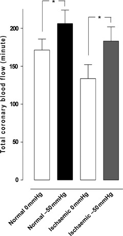
Several studies have suggested that mediastinitis is a strong predictor for poor long-term survival after coronary artery bypass surgery (CABG). In those studies, several conventional wound-healing techniques were used. Previously, we have shown no difference in long-term survival between CABG patients with topical negative pressure (TNP)-treated mediastinitis and CABG patients without mediastinitis. The present study was designed to elucidate if TNP, applied over the myocardium, resulted in an increase of the total amount of coronary blood flow. Six pigs underwent median sternotomy. The coronary blood flow was measured, before and after the application of TNP (-50 mmHg), using coronary electromagnetic flow meter probes. Analyses were performed before left anterior descending artery (LAD) occlusion (normal myocardium) and after 20 minutes of LAD occlusion (ischaemic myocardium). Normal myocardium: 171.3 +/- 14.5 ml/minute before to 206.3 +/- 17.6 ml/minute after TNP application, P < 0.05. Ischaemic myocardium: 133.7 +/- 18.4 ml/minute before to 183.2 +/- 18.9 ml/minute after TNP application, P < 0.05. TNP of -50 mmHg applied over the LAD region induced a significant increase in the total coronary blood flow in both normal and ischaemic myocardium.
Figures
Similar articles
-
A compare between myocardial topical negative pressure levels of -25 mmHg and -50 mmHg in a porcine model.BMC Cardiovasc Disord. 2008 Jun 22;8:14. doi: 10.1186/1471-2261-8-14. BMC Cardiovasc Disord. 2008. PMID: 18570679 Free PMC article.
-
No hypoperfusion is produced in the epicardium during application of myocardial topical negative pressure in a porcine model.J Cardiothorac Surg. 2007 Dec 6;2:53. doi: 10.1186/1749-8090-2-53. J Cardiothorac Surg. 2007. PMID: 18062803 Free PMC article.
-
Impact of different topical negative pressure levels on myocardial microvascular blood flow.Cardiovasc Revasc Med. 2008 Jan-Mar;9(1):29-35. doi: 10.1016/j.carrev.2007.09.001. Cardiovasc Revasc Med. 2008. PMID: 18206635
-
Topical negative pressure wound therapy: a review of its role and guidelines for its use in the management of acute wounds.Int Wound J. 2008 Oct;5(4):511-29. doi: 10.1111/j.1742-481X.2008.00437.x. Epub 2008 Sep 19. Int Wound J. 2008. PMID: 18808432 Free PMC article. Review.
-
[Sternal osteitis and mediastinitis after coronary artery bypass graft surgery].Ann Chir. 1991;45(2):128-35. Ann Chir. 1991. PMID: 2018332 Review. French.
Cited by
-
Influence of negative-pressure wound therapy on tissue oxygenation of the foot.Arch Plast Surg. 2014 Nov;41(6):668-72. doi: 10.5999/aps.2014.41.6.668. Epub 2014 Nov 3. Arch Plast Surg. 2014. PMID: 25396178 Free PMC article.
-
Prevention, Diagnosis and Management of Post-Surgical Mediastinitis in Adults Consensus Guidelines of the Spanish Society of Cardiovascular Infections (SEICAV), the Spanish Society of Thoracic and Cardiovascular Surgery (SECTCV) and the Biomedical Research Centre Network for Respiratory Diseases (CIBERES).J Clin Med. 2021 Nov 26;10(23):5566. doi: 10.3390/jcm10235566. J Clin Med. 2021. PMID: 34884268 Free PMC article. Review.
-
Negative pressure wound therapy for patients with mediastinitis: A meta-analysis.Int Wound J. 2020 Dec;17(6):2019-2025. doi: 10.1111/iwj.13494. Epub 2020 Aug 27. Int Wound J. 2020. Retraction in: Int Wound J. 2025 Apr;22(4):e70646. doi: 10.1111/iwj.70646. PMID: 32856392 Free PMC article. Retracted.
References
-
- Morykwas MJ, Argenta LC, Shelton‐Brown EI, McGuirt W. Vacuum‐assisted closure: a new method for wound control and treatment: animal studies and basic foundation. Ann Plast Surg 1997;38:553–62. - PubMed
-
- Argenta LC, Morykwas MJ. Vacuum‐assisted closure: a new method for wound control and treatment: clinical experience. Ann Plast Surg 1997;38:563–76; discussion 77. - PubMed
-
- Armstrong DG, Lavery LA. Negative pressure wound therapy after partial diabetic foot amputation: a multicentre, randomised controlled trial. Lancet 2005;366:1704–10. - PubMed
-
- O’Connor J, Kells A, Henry S, Scalea T. Vacuum‐assisted closure for the treatment of complex chest wounds. Ann Thorac Surg 2005;79:1196–200. - PubMed
-
- Brown KM, Harper FV, Aston WJ, O’Keefe PA, Cameron CR. Vacuum‐assisted closure in the treatment of a 9‐year‐old child with severe and multiple dog bite injuries of the thorax. Ann Thorac Surg 2001;72:1409–10. - PubMed
Publication types
MeSH terms
LinkOut - more resources
Full Text Sources
Medical
