. 2009 Jan;71(1):35-47.
doi: 10.1111/j.1365-2958.2008.06503.x. Epub 2008 Nov 7.

The Fusobacterium nucleatum outer membrane protein RadD is an arginine-inhibitable adhesin required for inter-species adherence and the structured architecture of multispecies biofilm

Affiliations

The Fusobacterium nucleatum outer membrane protein RadD is an arginine-inhibitable adhesin required for inter-species adherence and the structured architecture of multispecies biofilm

Christopher W Kaplan et al. Mol Microbiol. 2009 Jan.

Abstract

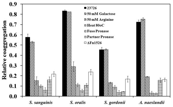
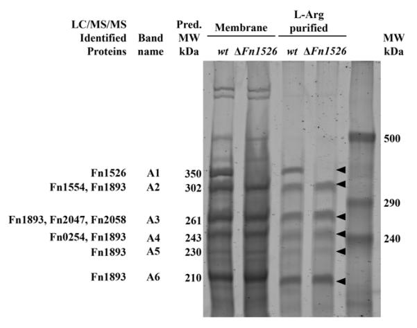
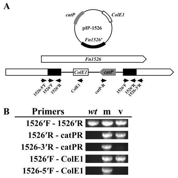
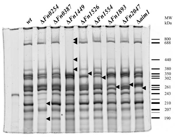
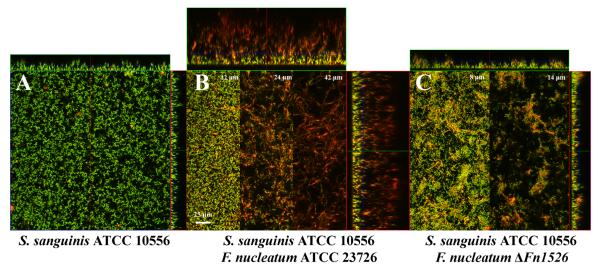
A defining characteristic of the suspected periodontal pathogen Fusobacterium nucleatum is its ability to adhere to a plethora of oral bacteria. This distinguishing feature is suggested to play an important role in oral biofilm formation and pathogenesis, with fusobacteria proposed to serve as central 'bridging organisms' in the architecture of the oral biofilm bringing together species which would not interact otherwise. Previous studies indicate that these bacterial interactions are mediated by galactose- or arginine-inhibitable adhesins although genetic evidence for the role and nature of these proposed adhesins remains elusive. To characterize these adhesins at the molecular level, the genetically transformable F. nucleatum strain ATCC 23726 was screened for adherence properties, and arginine-inhibitable adhesion was evident, while galactose-inhibitable adhesion was not detected. Six potential arginine-binding proteins were isolated from the membrane fraction of F. nucleatum ATCC 23726 and identified via mass spectroscopy as members of the outer membrane family of proteins in F. nucleatum. Inactivation of the genes encoding these six candidates for arginine-inhibitable adhesion and two additional homologues revealed that only a mutant derivative carrying an insertion in Fn1526 (now designated as radD) demonstrated significantly decreased co-aggregation with representatives of the gram-positive 'early oral colonizers'. Lack of the 350 kDa outer membrane protein encoded by radD resulted in the failure to form the extensive structured biofilm observed with the parent strain when grown in the presence of Streptococcus sanguinis ATCC 10556. These findings indicate that radD is responsible for arginine-inhibitable adherence of F. nucleatum and provides definitive molecular evidence that F. nucleatum adhesins play a vital role in inter-species adherence and multispecies biofilm formation.


Figures

Fig. 1
Fig. 1
Spectrophotometric co-aggregation assay of F. nucleatum ATCC 23726 and Gram-positive partner strains. Wild-type F. nucleatum ATCC 23726 co-aggregated with each of the Gram-positive partner strains tested. Treatment with heat (80°C, 15 min.) and pronase (10 mg ml-1, 2 h) resulted in approximately 90% loss of co-aggregation in both partner strains. Addition of arginine inhibited co-aggregation with each strain, while galactose had no effect on co-aggregation. Disruption of the gene encoding Fn1526 resulted in a loss of co-aggregation comparable to the effect seen with the addition of arginine. Relative co-aggregation was determined by dividing the difference between the total turbidity of each partner strain and the co-aggregation supernatant turbidity by the total turbidity of each partner strain. The data presented are based on experiments conducted in triplicate. All co-aggregation values except for galactose inhibition (P = 0.01) were significantly different from wild-type (P >0.0001).
Fig. 2
Fig. 2
Purified membrane proteins and arginine binding proteins from F. nucleatum ATCC 23726 wild-type and ΔFn1526 strains analyzed by 4% SDS-PAGE. Six arginine binding proteins were isolated from the wild-type strain and identified by LC/MS/MS. Gene inactivation of Fn1526 resulted in the absence of a 350 kDa protein in the membrane preparations from the Fn1526 mutant. The ‘Fn’ designation indicates the ORFs annotated in the F. nucleatum ATCC 25586 genome. Arrows indicate the six bands (A1-A6) that were analyzed by LC/MS/MS.
Fig. 3
Fig. 3
Analysis of the ΔFn1526 gene inactivation mutant. A. Organization of Fn1526 with insertion of the inactivation plasmid pIP-1526. Black arrows indicate the location of primers used for construction of the mutant and the analysis. Gene Fn1526 was shown as an example of the methodology for analyzing each insertion. B. Confirmation of plasmid insertion into Fn1526 by PCR analysis of the ΔFn1526 mutant (m) with wild-type (wt) and pIP-1526 vector (v) as controls. Fragments were amplified from the mutant strain, but not the controls, when using primer pairs where one was outside the insertion site (Fn1526-5′F/ColE1 and Fn1526-3′R/catPR).
Fig. 4
Fig. 4
Purified membrane proteins from eight OMP mutant strains analyzed by 4% SDS-PAGE. Arrows indicate protein bands missing in each gene inactivation mutant. The Fn1526 and four Fn1449 bands contained peptides for their respective proteins when analyzed by LC/MS/MS. The schematic to right represents each observed band in the wild-type strain with the relative molecular mass of the bands missing in the mutants indicated.
Fig. 5
Fig. 5
Typical biofilm growth after 48 h visualized by CSLM. Biofilms were stained with HEX-labeled F. nucleatum specific probes (red) and counterstained with STYO9 (green). A. Monoculture of S. sanguinis ATCC 10556. B. Co-culture of F. nucleatum ATCC 23726 and S. sanguinis ATCC 10556 produced an 80-μm-thick biofilm. Sections from 12, 24 and 42 μm above the growth surface demonstrate the variation in the biofilm morphology as the height of the biofilm increases. C. Co-culture of F. nucleatum ΔFn1526 with S. sanguinis ATCC 10556. Sections from 8, and 14 μm above the growth surface demonstrate the variation in the biofilm morphology as the height of the biofilm increases.
Fig. 6
Fig. 6
Analysis of the Fn1526 operon in F. nucleatum ATCC 23726. The operon in F. nucleatum ATCC 25586 is organized with three genes upstream of Fn1526. Amplification of fragments from ATCC 23726 genomic DNA and cDNA using primers in Fn1529 (Fn1526orfF) and Fn1526 (Fn1526-5′R) indicate that the organization of the genes in ATCC 23726 is the same at that of ATCC 25586, and that Fn1526 to Fn1529 are transcribed as a single unit. No fragments were amplified using primers spanning gene Fn1525 (1525R) and Fn1526 (Fn1526-3′F). RNA was used as a negative control for genomic DNA contamination. Black arrowheads indicate the position of primers used in this analysis.

References

    1. Charbonneau M-E, Mourez M. Functional organization of the autotransporter adhesin involved in diffuse adherence. J. Bacteriol. 2007;189:9020–9029. - PMC - PubMed
    1. Cisar JO, Kolenbrander PE, McIntire FC. Specificity of co-aggregation reactions between human oral streptococci and strains of Actinomyces viscosus or Actinomyces naeslundii. Infect Immun. 1979;24:742–752. - PMC - PubMed
    1. Clark WB, Beem JE, Nesbitt WE, Cisar JO, Tseng CC, Levine MJ. Pellicle receptors for Actinomyces viscosus type 1 fimbriae in vitro. Infect. Immun. 1989;57:3003–3008. - PMC - PubMed
    1. Edwards AM, Grossman TJ, Rudney JD. Association of a high-molecular weight arginine-binding protein of Fusobacterium nucleatum ATCC 10953 with adhesion to secretory immunoglobulin A and co-aggregation with Streptococcus cristatus. Oral Microbiology and Immunology. 2007;22:217–224. - PubMed
    1. Haake SK, Yoder SC, Attarian G, Podkaminer K. Native plasmids of Fusobacterium nucleatum: characterization and use in development of genetic systems. J Bacteriol. 2000;182:1176–1180. - PMC - PubMed

Publication types

LinkOut - more resources