TBP domain symmetry in basal and activated archaeal transcription
- PMID: 19007415
- PMCID: PMC2670189
- DOI: 10.1111/j.1365-2958.2008.06512.x
TBP domain symmetry in basal and activated archaeal transcription
Abstract
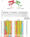
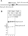
The TATA box binding protein (TBP) is the platform for assembly of archaeal and eukaryotic transcription preinitiation complexes. Ancestral gene duplication and fusion events have produced the saddle-shaped TBP molecule, with its two direct-repeat subdomains and pseudo-two-fold symmetry. Collectively, eukaryotic TBPs have diverged from their present-day archaeal counterparts, which remain highly symmetrical. The similarity of the N- and C-halves of archaeal TBPs is especially pronounced in the Methanococcales and Thermoplasmatales, including complete conservation of their N- and C-terminal stirrups; along with helix H'1, the C-terminal stirrup of TBP forms the main interface with TFB/TFIIB. Here, we show that, in stark contrast to its eukaryotic counterparts, multiple substitutions in the C-terminal stirrup of Methanocaldococcus jannaschii (Mja) TBP do not completely abrogate basal transcription. Using DNA affinity cleavage, we show that, by assembling TFB through its conserved N-terminal stirrup, Mja TBP is in effect ambidextrous with regard to basal transcription. In contrast, substitutions in either its N- or the C-terminal stirrup abrogate activated transcription in response to the Lrp-family transcriptional activator Ptr2.
Figures


Similar articles
-
Promoter architecture and response to a positive regulator of archaeal transcription.Mol Microbiol. 2005 May;56(3):625-37. doi: 10.1111/j.1365-2958.2005.04563.x. Mol Microbiol. 2005. PMID: 15819620
-
Hybrid Ptr2-like activators of archaeal transcription.Mol Microbiol. 2009 Nov;74(3):582-93. doi: 10.1111/j.1365-2958.2009.06884.x. Epub 2009 Sep 22. Mol Microbiol. 2009. PMID: 19775246
-
The basal transcription factors TBP and TFB from the mesophilic archaeon Methanosarcina mazeii: structure and conformational changes upon interaction with stress-gene promoters.J Mol Biol. 2001 Jun 8;309(3):589-603. doi: 10.1006/jmbi.2001.4705. J Mol Biol. 2001. PMID: 11397082
-
Archaeal chromatin and transcription.Mol Microbiol. 2003 May;48(3):587-98. doi: 10.1046/j.1365-2958.2003.03439.x. Mol Microbiol. 2003. PMID: 12694606 Review.
-
Role of the TATA-box binding protein (TBP) and associated family members in transcription regulation.Gene. 2022 Jul 30;833:146581. doi: 10.1016/j.gene.2022.146581. Epub 2022 May 18. Gene. 2022. PMID: 35597524 Review.
Cited by
-
The neomuran revolution and phagotrophic origin of eukaryotes and cilia in the light of intracellular coevolution and a revised tree of life.Cold Spring Harb Perspect Biol. 2014 Sep 2;6(9):a016006. doi: 10.1101/cshperspect.a016006. Cold Spring Harb Perspect Biol. 2014. PMID: 25183828 Free PMC article. Review.
-
Displacement of the transcription factor B reader domain during transcription initiation.Nucleic Acids Res. 2018 Nov 2;46(19):10066-10081. doi: 10.1093/nar/gky699. Nucleic Acids Res. 2018. PMID: 30102372 Free PMC article.
-
Archaeal transcription.Transcription. 2020 Oct;11(5):199-210. doi: 10.1080/21541264.2020.1838865. Epub 2020 Oct 28. Transcription. 2020. PMID: 33112729 Free PMC article. Review.
-
Archaeal histone-based chromatin structures regulate transcription elongation rates.Commun Biol. 2024 Feb 27;7(1):236. doi: 10.1038/s42003-024-05928-w. Commun Biol. 2024. PMID: 38413771 Free PMC article.
-
The Lrp family of transcription regulators in archaea.Archaea. 2010 Nov 30;2010:750457. doi: 10.1155/2010/750457. Archaea. 2010. PMID: 21151646 Free PMC article.
References
-
- Bell SD, Jackson SP. Mechanism and regulation of transcription in archaea. Curr Opin Microbiol. 2001;4:208–213. - PubMed
-
- Bryant GO, Martel LS, Burley SK, Berk AJ. Radical mutations reveal TATA-box binding protein surfaces required for activated transcription in vivo. Genes Dev. 1996;10:2491–2504. - PubMed
-
- Burley SK. The TATA box binding protein. Curr Opin Struct Biol. 1996;6:69–75. - PubMed
-
- Chen CH, Sigman DS. Chemical conversion of a DNA-binding protein into a site-specific nuclease. Science. 1987;237:1197–1201. - PubMed
Publication types
MeSH terms
Substances
Grants and funding
LinkOut - more resources
Full Text Sources
Miscellaneous

