. 2008 Dec;36(12):1714-27.
doi: 10.1016/j.exphem.2008.08.010.

Dynamin 3 participates in the growth and development of megakaryocytes

Affiliations

Dynamin 3 participates in the growth and development of megakaryocytes

Jo-Anna Reems et al. Exp Hematol. 2008 Dec.

Abstract

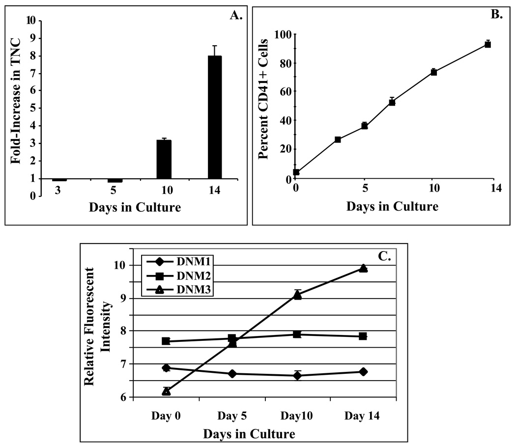
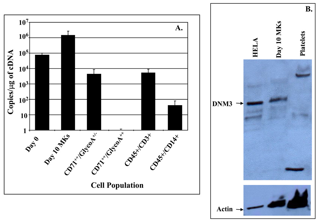
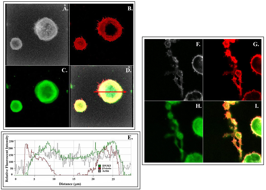
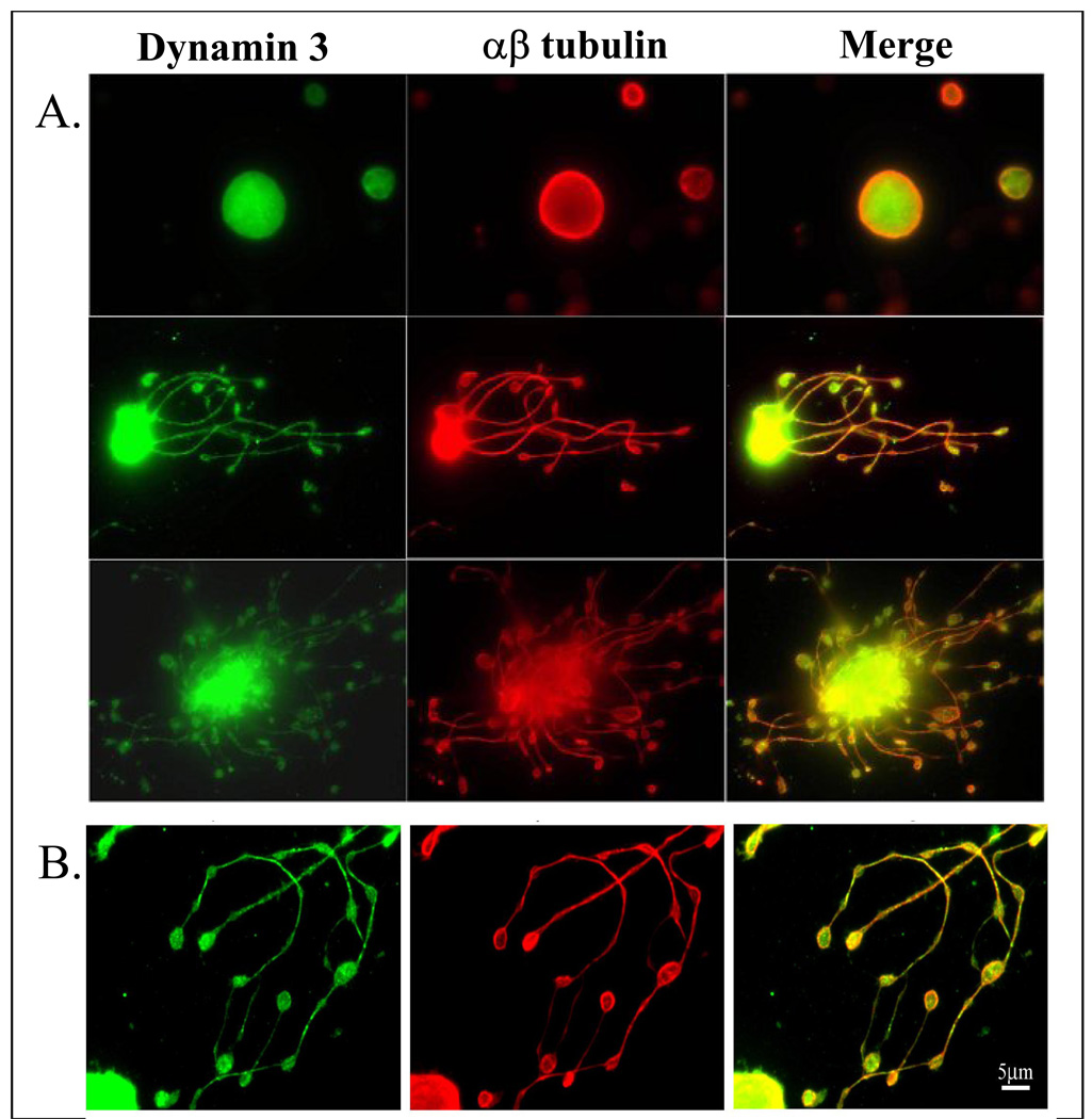
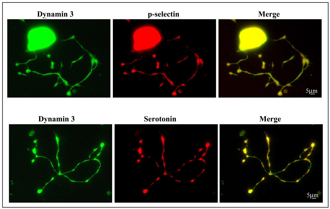
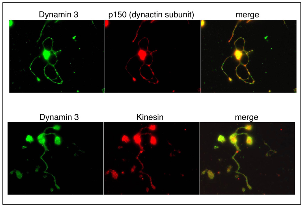
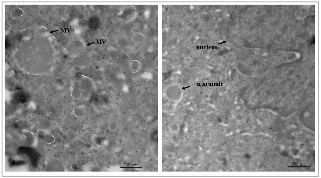
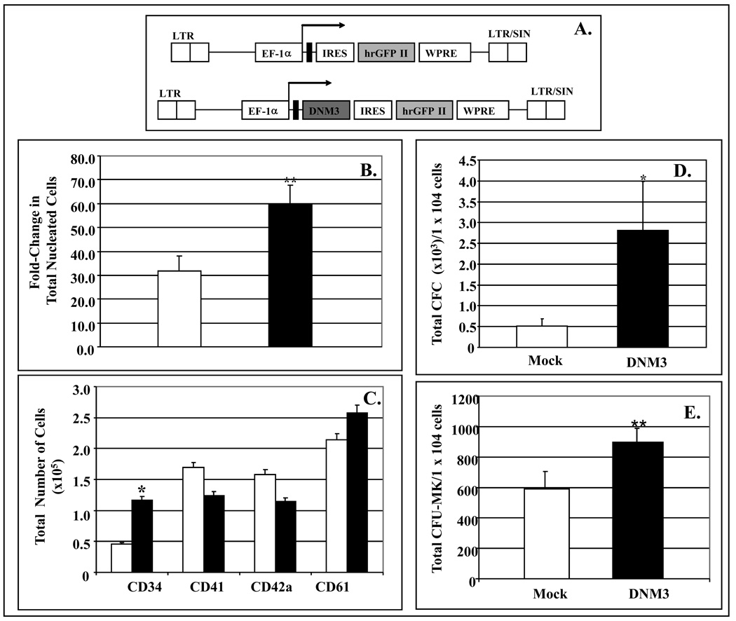
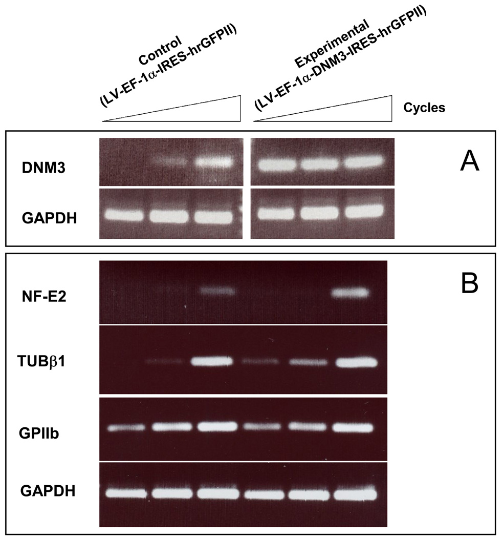
High-density oligonucleotide microarrays were used to compare gene expression profiles from uncultured CD34+/CD38lo cells and culture-derived megakaryocytes (MKs). As previously published, three replicate microarray data sets from three different sources of organ donor marrow were analyzed using the software program Rosetta Resolver. After setting a stringent p value of <or=0.001 with a fold change cutoff of three or more in expression level, dynamin 3 (DNM3) was identified to be differentially expressed during the course of MK development with a mean fold-change of 8.2+/-2.1 (mean+/-standard deviation). DNM3 is a member of a family of mechanochemical enzymes (DNM1, DNM2, and DNM3) known for their participation in membrane dynamics by hydrolyzing nucleotides to link cellular membranes to the actin cytoskeleton. Real-time quantitative polymerase chain reaction confirmed that DNM3 increased by 20.7-+/-3.4-fold (n=4, p=0.09) during megakaryocytopoiesis and Western blot analysis showed that DNM3 protein was expressed in human MKs. Confocal microscopy revealed that DNM3 was distributed diffusely throughout the cytoplasm of MKs with a punctate appearance in proplatelet processes. Immunogold electron microscopy also showed that DNM3 is widely distributed in the cytoplasm of MKs, with no apparent localization to specific organelles. The open reading frame of DNM3 was cloned from culture-derived human MKs and determined to be 100% identical to the protein encoded by the DNM3 transcript variant ENST00000367731 published in the Ensemble database. Overexpression of DNM3 in umbilical cord blood CD34+ cells resulted in an increase in total nucleated cells, an amplification of total colony-forming cells and colony-forming unit-megakaryocytes, and a concomitant increase in the expression of nuclear factor erythroid 2 (NF-E2) and beta-tubulin. Together these findings provide the first evidence that a member of the dynamin family of mechanochemical enzymes is present in human MKs and indicate that DNM3 is an excellent candidate for playing an important role in mediating cytoskeleton and membrane changes that occur during MK/platelet development.


Figures

Figure 1
Figure 1. DNM3 gene expression increases during megakaryocyte development
Cultures were initiated with purified human CD34+/CD38lo cells and maintained in media supplemented with IL3, IL6, SCF and Tpo. A) Fold increase in total nucleated cells at 0, 3, 5 10 and 14 days of culture. B) Percent of total cells expressing the CD41 antigen at indicated time points C) Relative fluorescent intensities from a single experiment for dynamins 1, 2 and 3 as detected on Affymetrix Gene Chip U133A \. Transcript levels for all three dynamins were determined from uncultured CD34+/CD38lo cells (Day 0) and their progeny cells harvested on days 5, 10 and 14 of culture in serum deprived media supplemented with IL3, IL6, SCF, and Tpo.
Figure 2
Figure 2. Quantitative RT-PCR (qRT-PCR) and Western Blot analysis of DNM3
A) Cell populations tested by qRT-PCR include: Day 0 (i.e. uncultured CD34+/CD38lo BM cells); Day 10 MKs (i.e. culture derived MKs generated from CD34+/CD38lo after 10 days of culture with IL3, IL6, SCF & Tpo); CD71++/GlycoA+/− and CD71++/GlycoA+/− (i.e. isolated from volunteer donor marrow mononuclear fractions); and CD45+/CD3+, and CD45+/CD14+ cells (i.e. isolated from organ donor marrow);. Data are shown as mean±SD. B)Western Blot analysis was performed on Day 10 culture derived human MKs produced from umbilical cord blood CD34+ cells cultured for 10 days in media supplemented with IL3, IL6, SCF and Tpo. After 10 days of culture, the cells were harvested and stained with anti-CD41-PE. The stained cells were sorted by flow cytometry to obtain purified MKs, which were used to prepare protein lysates. Platelets were obtained from human peripheral blood. HeLa cell lysates were purchased and used as a positive control.
Figure 3
Figure 3. Representative immunofluorescent confocal micrographs showing the distribution of DNM3 relative to tubulin α/β and actin in representative culture derived human MKs and MKs displaying proplatelet processes produced from human UCB CD34+ cell
(A–D) Staining patterns in a culture derived human MK produced from umbilical cord blood CD34+ cells. Shown separately in panels are: A) Bodipy-phalloidin (white), B.) anti-DNM3 (green) and C) anti-tubulin (red). D) Merge of actin, DNM3 and tubulin staining. Arrow represents the cross-sectional region that was scanned to obtain the histogram in 3E, which graphically profiles actin (gray line) DNM3 (green line) and tubulin (red line). (F–I) Representative proplatelet processes stained with the following: F) Bodipy phalloidin (white), G.) anti-DNM3 (green) and H) anti-tubulin (red). I) Merge of actin, DNM3 and tubulin staining.
Figure 4
Figure 4. Representative immunofluorescent confocal micrographs showing the distribution of DNM3 relative to αβtubulin in a murine MK displaying proplatelet processes. Magnifications: A) 90x; B) 150x
Figure 5
Figure 5. Representative immunofluorescent confocal micrographs showing the distribution of DNM3 relative to the expression of p-selectin and serotonin. Magnification 90X
Figure 6
Figure 6. Representative immunofluorescent confocal micrographs showing the distribution of DNM3 relative to the motor proteins dynactin and kinesin. Magnification 90X
Figure 7
Figure 7. Transmission electron micrograph showing DNM3 immunogold labeling in a murine megakaryocyte
Figure 8
Figure 8. Response of umbilical cord blood CD34+ cells infected with a lentiviral vector expressing DNM3
UCB CD34+ cells were infected with control vector (white bars) or test vector to overexpress DNM3 (black bars). A) Diagram of the control lentiviral vector, pLV-EF-1α-IRES-hrGFP II and the DNM3 vector pLV-EF-1α-DNM3-IRES-hrGFP II. The elongation factor 1-alfa promoter (EF-1α) was used to drive expression of the hrGFP II in the control vector and DNM3 in the experimental vector. Abbreviations: LTR, long terminal repeat; EF-1a, elongation factor 1-alfa promoter: SIN, self-inactivating; IRES, internal ribosome binding site; hrGFPII, humanized R. reniformis green fluorescent protein; WPRE, the post-transcriptional regulatory element of the woodchuck hepatitis virus to enhance transgene expression; pA, polyadenylation signal B) Significant fold-increase in the total number of nucleated cells generated after 10 days of culture (**p<0.001). C) Absolute number cell expressing the indicated cell surface markers (*p<0.05D) Total number of CFC per 1×10⁴ progeny cells plated. E) Total number of CFU-MK per 1×10⁴ progeny cells plated.
Figure 9
Figure 9. Semi-quantitative RT-PCR analysis of RNA extracted from umbilical cord blood CD34+ cells infected with a DNM3 expressing lentivirus (LV-EF1-α-DNM3-IRES-hrGFPII) or mock infected (LV-EF1-α-IRES-hrGFPII)
Ethidium bromide-stained agarose gels of RT-PCR products differing by 3-cycle increments (37, 40, 43). GAPDH was used as a housekeeping gene control. A) Changes in mRNA expression levels for DNM3. B) Changes in expression levels for NF-E2, β1tubulin and GPIIb.

Similar articles

Cited by

References

    1. Orth JD, McNiven MA. Dynamin at the actin-membrane interface [review] Curr Opin Cell Biol. 2003;15:31–39. - PubMed
    1. Shpetner HS, Vallee RB. Identification of dynamin, a novel mechanochemical enzyme that mediates interactions between microtubules. Cell. 1989;59:421–432. - PubMed
    1. Ochoa GC, Slepnev VI, Neff L, et al. A functional link between dynamin and the actin cytoskeleton at podosomes. J Cell Biol. 2000;150:377–389. - PMC - PubMed
    1. McNiven MA, Kim L, Krueger EW, Orth JD, Cao H, Wong TW. Regulated interactions between dynamin and the actin-binding protein cortactin modulate cell shape. J Cell Biol. 2000;151:187–198. - PMC - PubMed
    1. Krutchen AE, McNiven MA. Dynamin as a mover and pincher during cell migration and invasion. J Cell Sci. 2006;119:1683–1690. - PubMed

Publication types

MeSH terms