. 2009 Jan 1;325(1):162-70.
doi: 10.1016/j.ydbio.2008.10.023. Epub 2008 Oct 31.

Rac1 deficiency in the forebrain results in neural progenitor reduction and microcephaly

Affiliations

Rac1 deficiency in the forebrain results in neural progenitor reduction and microcephaly

Lei Chen et al. Dev Biol. .

Abstract

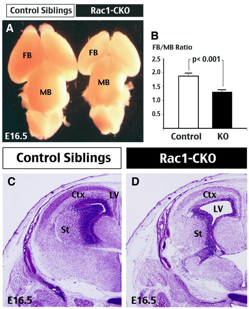
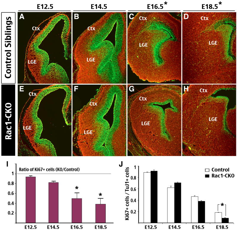
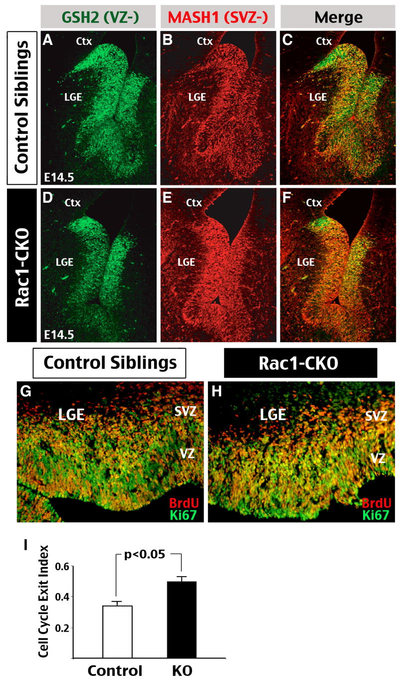
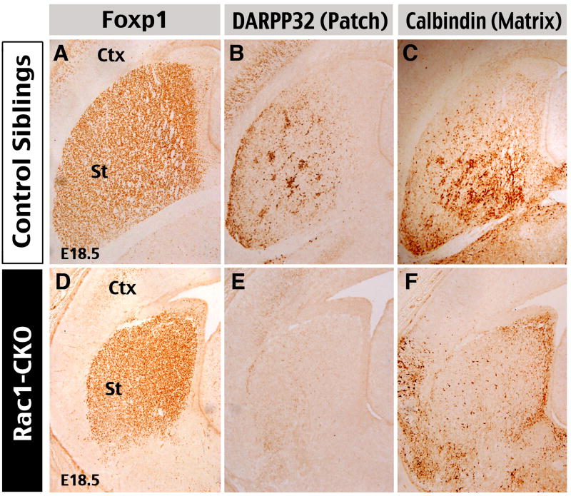
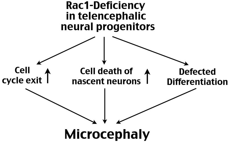
The Rho family of small GTPases has been implicated in many neurological disorders including mental retardation, but whether they are involved in primary microcephaly (microcephalia vera) is unknown. Here, we examine the role of Rac1 in mammalian neural progenitors and forebrain development by a conditional gene-targeting strategy using the Foxg1-Cre line to delete floxed-Rac1 alleles in the telencephalic ventricular zone (VZ) of mouse embryos. We found that Rac1 deletion in the telencephalic VZ progenitors resulted in reduced sizes of both the striatum and cerebral cortex. Analyses further indicated that this abnormality was caused by accelerated cell-cycle exit and increased apoptosis during early corticogenesis (approximately E14.5), leading to a decrease of the neural progenitor pool in mid-to-late telencephalic development (E16.5 to E18.5). Moreover, the formation of patch-matrix compartments in the striatum was impaired by Rac1-deficiency. Together, these results suggest that Rac1 regulates self-renewal, survival, and differentiation of telencephalic neural progenitors, and that dysfunctions of Rac1 may lead to primary microcephaly.


Figures

Figure 1
Figure 1. Deletion of Rac1 in forebrain neural progenitors results in microcephaly
(A) Gross brain morphology of E16.5 Rac1-CKO (Foxg1-Cre: Rac1-flox/flox) embryos and control siblings (Rac1-flox/+). The Rac1-CKO brains have a disproportional smaller forebrain (FB) than control siblings. (B) Three dimensional widths and lengths of the FB and midbrain (MB) were measured in control and Rac1-CKO embryos for comparison. Data were compiled from 6 sets of matched embryos from different litters. p < 0.001 was determined by t-test. (C, D) Nissl stain of coronal sections at E16.5 revealed a diminished size of the cerebral cortex (Ctx) and striatum (St) in Rac1-CKO embryos (D), resulting in an enlarged lateral ventricle (LV), when compared to those in the control embryos (C).
Figure 2
Figure 2. Forebrain-Rac1 deficiency results in gradual reduction of the progenitor pool
(A-H) Double-immunofluorescence staining of Ki67 (Green; a marker for proliferative cells) and Tuj1 (Red; a neuronal marker) was performed in E12.5 to E18.5 forebrains of control (A-D) and Rac1-CKO (E-H) embryos. Note the reduction of Ki67-stained domains in E16.5 and E18.5 Rac1-CKO embryos (asterisks). (I) The total numbers of Ki67 positive cells in comparable forebrain sections of control and Rac1-CKO embryos were counted from E12.5 to E18.5 for comparison. Data were collected from 3-4 embryos for each age. This analysis showed a significant reduction of Ki67-positive cells in Rac1-CKO embryos at E16.5 and E18.5 (asterisk: p < 0.01 by t-test). (J) The ratios of Ki67-positive cells over TuJ1-positive cells in the control and Rac1-CKO embryos from E12.5 to E18.5 developmental stages were quantified (asterisk: p < 0.01 by t-test).
Figure 3
Figure 3. Rac1 deficiency in neural progenitors accelerates cell cycle exit
(A-F) Double-immunofluorescence staining of GSH2 (Green; a ventral telencephalic neural progenitor marker) and Mash1 (Red; a marker for progenitors normally located between the ventricular and subventricualr zones of the ventral telencephalon) was performed in E14.5 control (A-C) and Rac1-CKO (D-F) embryos. Note the expansion of Mash1-positive domain and reciprocal reduction of GSH2-positive cells in the lateral ganglion eminence (LGE) of Rac1-CKO embryos. Shown is the representative result in 5 sets of embryos. (G, H) A cell cycle exit assay performed in Rac1-CKO (H) and control sibling (G) embryos by injecting BrdU to the pregnant mouse at the gestational 14.5 day followed by double anti-BrdU/Ki67 staining of the embryos collected 24 hours later. (I) The cell cycle exit index was calculated by counting those cells that are positive for BrdU but negative for Ki67 staining over the total number of BrdU-positive cells in the LGE. This analysis showed approximately 30% increase in the cell cycle exit index in Rac1-CKO, when compared to that in control embryos (Data were collected from 5 sets of embryos, p < 0.05 by t-test).
Figure 4
Figure 4. Forebrain-Rac1 deficiency leads to increased apoptosis of nascent neurons
(A, B) Immunocytochemistry showed an increase of active Caspase-3 staining in the forebrain of Rac1-CKO embryos than those in control sibling embryos from E14.5 to E18.5, with the difference most pronounced at E14.5. The majority of active Caspase-3-stained cells were located outside the ventricular zone (VZ), suggesting that they are nascent postmitotic cells. (C) The numbers of active Caspase-3-stained cells in comparable forebrain sections of control and Rac1-CKO embryos were counted from E14.5 to E18.5 for comparison. (Data were collected from 3 sets of embryos for each age, p < 0.01 by t-test).
Figure 5
Figure 5. Forebrain Rac1-deficiency causes abnormal differentiation of the striatum
The differentiation and compartmentalization of the striatum was examined in E18.5 embryos by immunostaining against Foxp1 (a marker for all striatal projection neurons), DARPP-32 (a marker of the patch compartment), and Calbindin (a matrix-compartment marker). This analysis showed that Rac1-CKO embryos have relatively normal expression of Foxp1 (D), but greatly decreased DARPP-32 expression (E) and ectopic distribution of Calbindin-positive cells (F) in the striatum, when compared to control embryos (A, B, C). Shown are typical staining patterns in five sets of embryos.
Figure 6
Figure 6. Abnormal forebrain development associated with Rac1 deficiency in neural progenitors
Loss of Rac1 in the telencephalic neural progenitors can accelerate cell cycle exit, increase apoptosis of nascent neurons, and impair neuronal differentiation at a late embryonic stage. The combination of these events may lead to a reduced size of the forebrain or microcephaly.

Similar articles

Cited by

References

    1. Abuelo D. Microcephaly syndromes. Semin Pediatr Neurol. 2007;14:118–127. - PubMed
    1. Benitah SA, Frye M, Glogauer M, Watt FM. Stem cell depletion through epidermal deletion of Rac1. Science. 2005;309:933–935. - PubMed
    1. Bond J, Scott S, Hampshire DJ, Springell K, Corry P, Abramowicz MJ, Mochida GH, Hennekam RC, Maher ER, Fryns JP, Alswaid A, Jafri H, Rashid Y, Mubaidin A, Walsh CA, Roberts E, Woods CG. Protein-truncating mutations in ASPM cause variable reduction in brain size. Am J Hum Genet. 2003;73:1170–7. - PMC - PubMed
    1. de la Rosa EJ, de Pablo F. Cell death in early neural development: Beyond the neurotrophic theory. Trends Neurosci. 2000;23:454–58. - PubMed
    1. Cecconi F, Alvarez-Bolado G, Meyer BI, Roth KA, Gruss P. Apaf1 (CED-4 homolog) regulates programmed cell death in mammalian development. Cell. 1998;94:727–37. - PubMed

MeSH terms