. 2009 Jan 16;385(2):457-68.
doi: 10.1016/j.jmb.2008.10.068. Epub 2008 Nov 5.

Essential role of PACT-mediated PKR activation in tunicamycin-induced apoptosis

Affiliations

Essential role of PACT-mediated PKR activation in tunicamycin-induced apoptosis

Madhurima Singh et al. J Mol Biol. .

Abstract

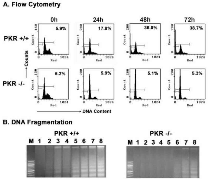
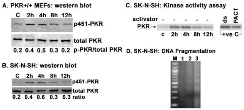
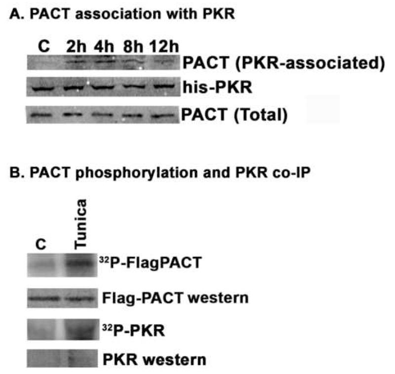
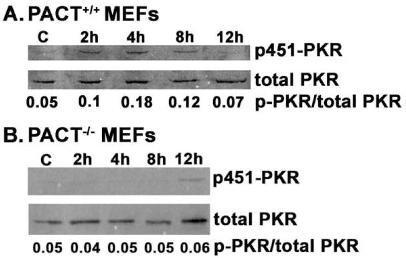
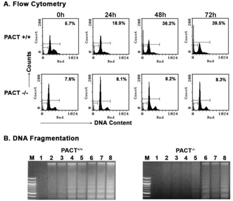
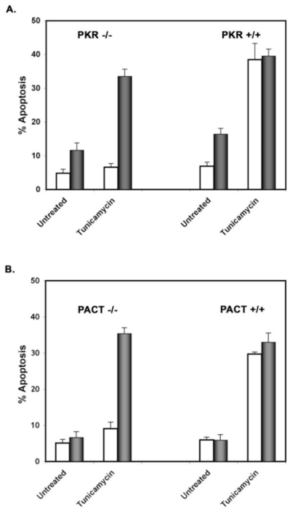
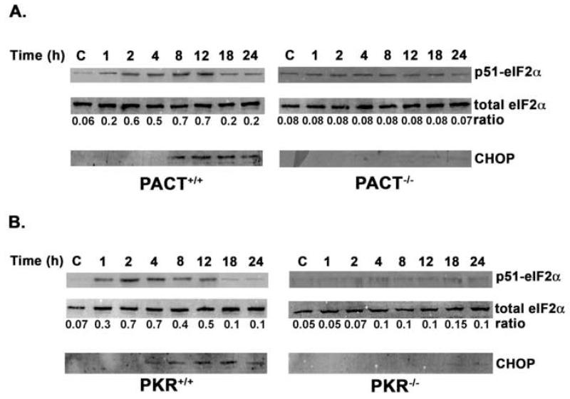
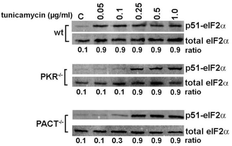
Cellular stresses such as disruption of calcium homeostasis, inhibition of protein glycosylation, and reduction of disulfide bonds result in accumulation of misfolded proteins in the endoplasmic reticulum (ER) and lead to cell death by apoptosis. Tunicamycin, which is an inhibitor of protein glycosylation, induces ER stress and apoptosis. In this study, we examined the involvement of double-stranded RNA (dsRNA)-activated protein kinase (PKR) and its protein activator PACT in tunicamycin-induced apoptosis. We demonstrate for the first time that PACT is phosphorylated in response to tunicamycin and is responsible for PKR activation by direct interaction. Furthermore, PACT-induced PKR activation is essential for tunicamycin-induced apoptosis, since PACT as well as PKR null cells are markedly resistant to tunicamycin and show defective eIF2alpha phosphorylation and C/EBP homologous protein (CHOP, also known as GADD153) induction especially at low concentrations of tunicamycin. Reconstitution of PKR and PACT expression in the null cells renders them sensitive to tunicamycin, thus demonstrating that PACT-induced PKR activation plays an essential function in induction of apoptosis.


Figures

Fig. 1
Fig. 1. PKR null MEFs are resistant to tunicamycin-induced apoptosis
A. Flow cytometry analysis of cell-cycle distribution after tunicamycin treatment. PKR+/+ and PKR−/− MEFs were treated with 0.1μg/ml tunicamycin. Cells were harvested at 24h, 48h, and 72h after the treatment and subjected to flow cytometry analysis after propidium iodide staining. The sub-G0/G1 cell population represents the apoptotic cells. The sub-G0/G1 percentages are displayed in each panel. These experiments were repeated twice, each time with duplicate samples. The most representative profiles from a single experiment are shown. B. DNA fragmentation analysis in response to tunicamycin. PKR+/+ and PKR−/− MEFs were treated with increasing concentrations of tunicamycin. 48 h after tunicamycin treatment, the fragmented DNA was analyzed. The cell type is as indicated on the top of the lanes. Lane 1, untreated cells; Lanes 2-8 cells treated with different concentrations of tunicamycin. Lane 2, 0.01μg/ml; lane 3, 0.025 μg/ml; lane 4, 0.05 μg/ml; lane 5, 0.1 μg/ml; lane 6, 0.25 μg/ml; lane 7, 0.5 μg/ml, and lane 8, 1 μg/ml. M: 100-bp ladder DNA size markers.
Fig. 2
Fig. 2. PKR is activated in both murine and human cells in response to tunicamycin
A. PKR+/+ MEFs: Western blot analysis with a phosphoserine-451 specific antibody. The MEFs were treated with 1 μg/ml tunicamycin and cell extracts were prepared at indicated times after the treatments. Phosphorylation of PKR was examined by western blot analysis with a phosphoserine-451 specific antibody. The same blot was stripped and re-probed with an anti-PKR monoclonal antibody to detect total PKR. The ratio of phosphorylated PKR/total PKR is represented below each lane. B. SK-N-SH: Western blot analysis with a phosphoserine-451 specific antibody. The SK-N-SH cells were treated with 1 μg/ml tunicamycin and cell extracts were prepared at indicated times after the treatments. Phosphorylation of PKR was examined by western blot analysis with a phosphoserine-451 specific antibody. The same blot was stripped and re-probed with an anti-PKR monoclonal antibody to detect total PKR. The ratio of phosphorylated PKR/total PKR is represented below each lane. C. SK-N-SH: PKR kinase activity assay. SK-N-SH cells were treated with 1 μg/ml tunicamycin and PKR activity was assayed from the cell extracts prepared at the indicated times. PKR was immunoprecipitated with a monoclonal antibody 71/10 and protein A-sepharose beads. PKR activity assay was performed with PKR attached to the protein A-sepharose beads without any exogenous activator added. Lanes marked +ve C are positive controls to show that activation occurs with extract from untreated cells with 0.1μg/ml polyI-polyC (dsRNA) and 0.2 pmoles of pure recombinant truncated PACT (amino acids 1-305) added to the kinase assay reaction in vitro. D. SK-N-SH: DNA fragmentation analysis in response to tunicamycin. SK-N-SH cells were treated with 1 μg/ml tunicamycin. At 24h and 48 h after tunicamycin treatment, the fragmented DNA was analyzed. Lane 1, untreated cells; Lanes 2, 24h treatment; and lane 3, 48h treatment. M: 100-bp ladder DNA size markers.
Fig. 3
Fig. 3. PACT activates PKR in response to tunicamycin
A. PACT associates with PKR in response to tunicamycin. SK-N-SH cells were treated with 1 μg/ml tunicamycin. The cells extracts were prepared at indicated times and 500 μg of protein from the cell extracts was allowed to interact with 1 μg of purified PKR protein immobilized on Ni-agarose beads. After washing the beads to remove unbound material, the PKR-associated PACT was analyzed by SDS-polyacrylamide gel electrophoresis followed by immunoblotting using anti-PACT antibody. The same blot was stripped and re-probed with anti-hexahistidine tag antibody to detect total PKR immobilized on Ni-agarose beads. To analyze total PACT amounts in the extracts, 100 μg of the total protein from extracts was analyzed by western blot analysis with anti-PACT antibody. B. PACT phosphorylation and PKR association in response to tunicamycin. SK-N-SH cells were transfected with flag-PACT/pCDNA3.1 (full-length/amino acids 1-313) to achieve overexpression of flag-PACT. 24h after transfection, the cells were treated with 1μg/ml tunicamycin for 4h in the presence of 0.5 mCi/ml 32P-orthophosphate in phosphate free medium. Phosphorylation status of PACT was examined by immunoprecipitation with anti-Flag monoclonal antibody followed by phosphorimager analysis (32P-FlagPACT). In the same gel, a weaker 32P-labeled band was observed at a position corresponding to PKR (32P-PKR). Aliquots of cell lysate with 100 μg total protein were also examined by western blot analysis for total flag-PACT with anti-flag antibody (Flag-PACT western). A western blot analysis was performed with anti-PKR monoclonal antibody on 1 g of cell extracts immunoprecipitated with anti-Flag M2 monoclonal antibody (PKR western). Subsequent autoradiographic analysis of the western blot also showed presence of 32P-labeling in the band corresponding to PKR.
Fig. 4
Fig. 4. PKR is not activated in PACT null MEFs
The PACT+/+ (A) and PACT−/− (B) MEFs were treated with 1 μg/ml tunicamycin and cell extracts were prepared at indicated times after the treatments. Phosphorylation of PKR was examined by western blot analysis with a phosphoserine-451 specific antibody. The same blot was stripped and re-probed with an anti-PKR monoclonal antibody to detect total PKR. The ratio of phosphorylated PKR/total PKR is represented below each lane.
Fig. 5
Fig. 5. PACT null MEFs are resistant to tunicamycin-induced apoptosis
A. Flow cytometry analysis of cell-cycle distribution after tunicamycin treatment. PACT+/+ and PACT−/− MEFs were treated with 0.1μg/ml tunicamycin. Cells were harvested at 24h, 48h, and 72h after the treatment and subjected to flow cytometry analysis as described. The sub-G0/G1 cell population represents the apoptotic cells. The sub-G0/G1 percentages are displayed in each panel. These experiments were repeated twice, each time with duplicate samples. The most representative profiles from a single experiment are shown. B. DNA fragmentation analysis in response to tunicamycin. PACT+/+ and PACT−/− MEFs were treated with increasing concentrations of tunicamycin. 48 h after tunicamycin treatment, the fragmented DNA was analyzed. The cell type is as indicated on the top of the lanes. Lane 1, untreated cells; Lanes 2-8 cells treated with different concentrations of tunicamycin. Lane 2, 0.01μg/ml; lane 3, 0.025 μg/ml; lane 4, 0.05 μg/ml; lane 5, 0.1 μg/ml; lane 6, 0.25 μg/ml; lane 7, 0.5 μg/ml, and lane 8, 1 μg/ml. M: 100-bp ladder DNA size markers.
Fig. 6
Fig. 6. Reconstitution of PKR and PACT expression restores the apoptotic response in null MEFs
A. Reconstitution of PKR expression. As indicated, PKR−/− or PKR+/+ MEFs grown on coverslips were transfected with 500 ng of pCDNA3.1 + 100 ng of pEGFPC1 (white bars) or 500 ng of flag-PKR/pCDNA3.1 + 100 ng of pEGFPC1 (black bars) using Effectene. The cells were observed for EGFP fluorescence 24 h after transfection and were treated with 0.1μg/ml of tunicamycin. The morphology of cells was monitored every 12 h. At 72 h after treatment, the cells were fixed and mounted in Vectashield with DAPI nuclear stain. White bars represent empty vector transfected cells and black bars represent cells with PKR expression construct transfection. At least 300 fluorescent (EGFP-positive) cells were counted as live or dead based on their morphology and nuclear condensation indicated by intense DAPI fluorescence. % apoptosis = (fluorescent dead cells with intense DAPI fluorescence/total fluorescent cells) × 100. B. Reconstitution of PACT expression. As indicated, PACT−/− or PACT+/+ MEFs grown on coverslips were transfected with 500 ng of pCDNA3.1 + 100 ng of pEGFPC1 (white bars) or 500 ng of flag-PACT/pCDNA3.1 (encoding full length PACT, amino acids 1-313) + 100 ng of pEGFPC1 (black bars) using Effectene. The cells were subjected to similar treatment and analysis as in A.
Fig. 7
Fig. 7. Functional requirement of PACT and PKR for eIF2α phosphorylation and CHOP expression in response to tunicamycin
A. eIF2α phosphorylation and CHOP induction in PACT null MEFs. PACT+/+ and PACT−/− MEFs were treated with 0.1 μg/ml tunicamycin. The cells extracts were prepared at indicated times and analyzed by western blot analysis. The same blot was stripped and re-probed with all antibodies. The numbers at the bottom of the panels represent the ratio of phosphorylated eIF2α to total eIF2α, calculated using the Imagequant software and Storm phosphorimager. The analysis was repeated five times and best representative blots are shown. The fold increases in ratios after tunicamycin treatment compared to the control samples were calculated at each time point and subjected to a statistical analysis with the use of the Student’s t test. The observed differences between +/+ and −/− MEFS are considered significant with all probability values being less than 0.05. B. eIF2α phosphorylation and CHOP induction in PKR null MEFs. PKR+/+ and PKR−/− MEFs were treated with 0.1 μg/ml tunicamycin. The cells extracts prepared at indicated times were analyzed as described in A.
Fig. 8
Fig. 8. Tunicamycin dose response
PKR+/+, PACT+/+, PKR−/−, and PACT−/− MEFs were treated with 0.05 μg/ml, 0.1 μg/ml, 0.25 μg/ml, 0.5 μg/ml, and 1.0 μg/ml tunicamycin as indicated on top of the lanes. The cells extracts were prepared at 2 h after the treatments and analyzed by western blot analysis. The same blot was stripped and re-probed with both the antibodies. The numbers at the bottom of the panels represent the ratio of phosphorylated eIF2α to total eIF2α, calculated using the Imagequant software and Storm phosphorimager. Since the results obtained from PKR+/+ and PACT +/+ MEFS were identical, only the PKR+/+ MEF results are represented as wt panel.

Similar articles

Cited by

References

    1. Meurs E, Chong K, Galabru J, Thomas NS, Kerr IM, Williams BR, Hovanessian AG. Molecular cloning and characterization of the human double-stranded RNA-activated protein kinase induced by interferon. Cell. 1990;62:379–390. - PubMed
    1. Williams BR. PKR; a sentinel kinase for cellular stress. Oncogene. 1999;18:6112–6120. - PubMed
    1. Garcia MA, Gil J, Ventoso I, Guerra S, Domingo E, Rivas C, Esteban M. Impact of protein kinase PKR in cell biology: from antiviral to antiproliferative action. Microbiol. Mol. Biol. Rev. 2006;70:1032–1060. - PMC - PubMed
    1. Hovanessian AG, Galabru J. The double-stranded RNA-dependent protein kinase is also activated by heparin. Eur. J. Biochem. 1987;167:467–473. - PubMed
    1. Sadler AJ, Williams BR. Structure and function of the protein kinase R. Curr. Top. Microbiol. Immunol. 2007;316:253–292. - PubMed

Publication types