

Differentiation-induced uroplakin III expression promotes urothelial cell death in response to uropathogenic E. coli
- PMID: 19007907
- PMCID: PMC2847841
- DOI: 10.1016/j.micinf.2008.10.008
Differentiation-induced uroplakin III expression promotes urothelial cell death in response to uropathogenic E. coli
Abstract
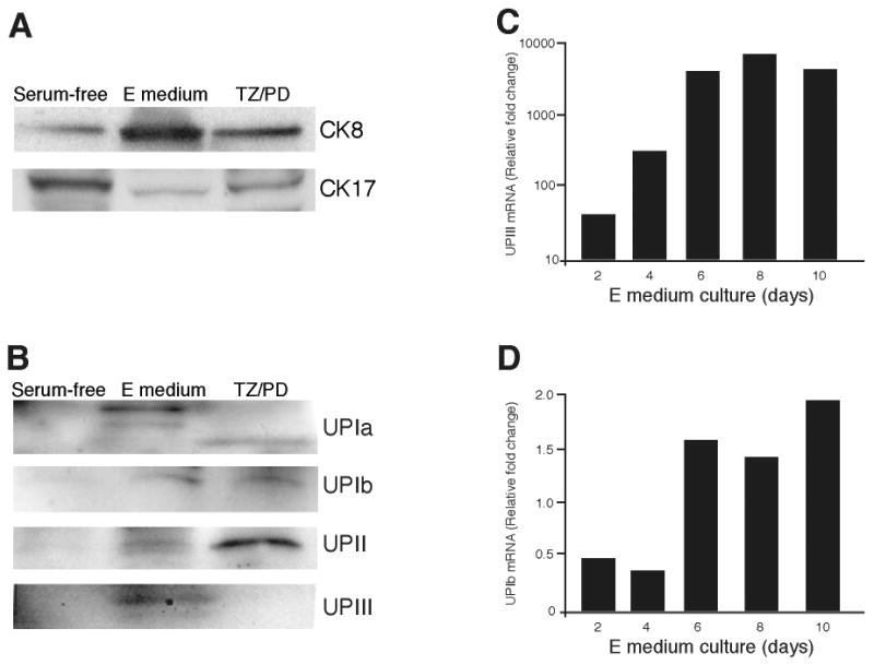
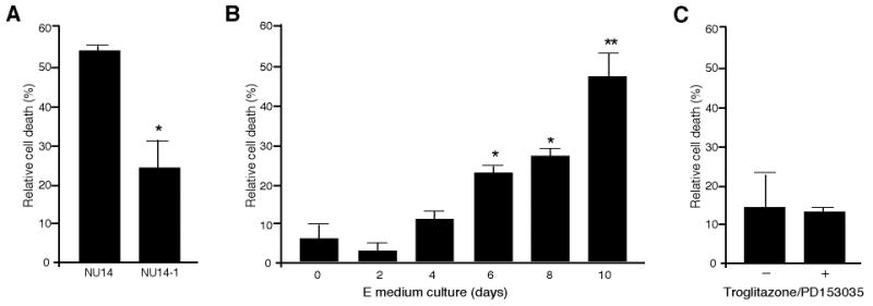
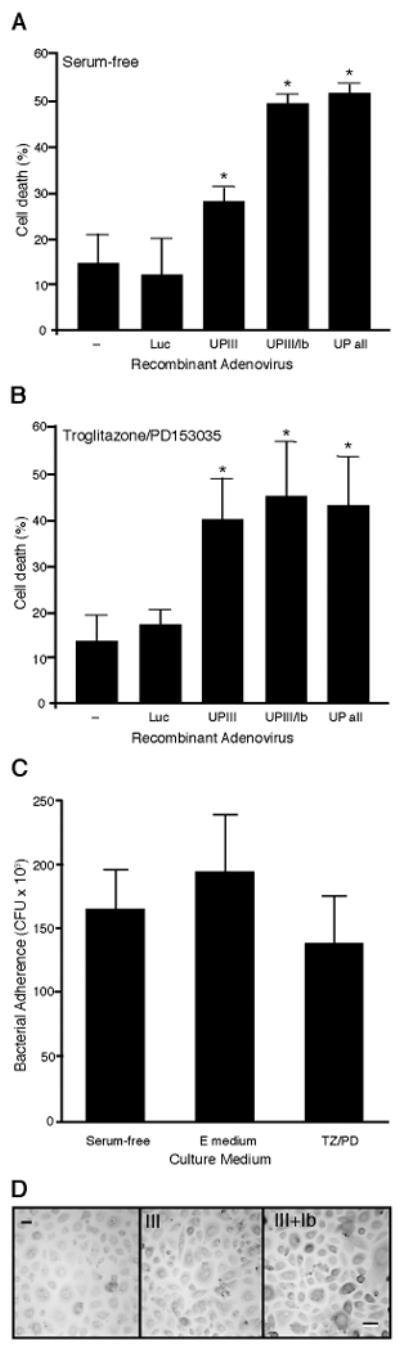
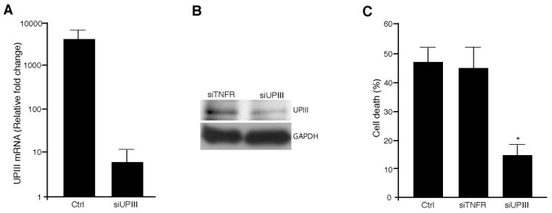
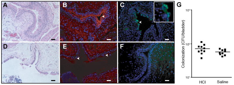
Uropathogenic E. coli (UPEC) expressing type 1 pili underlie most urinary tract infections (UTIs). UPEC adherence to the bladder urothelium induces a rapid apoptosis and exfoliation of terminally differentiated urothelial cells, a critical event in pathogenesis. Of the four major uroplakin proteins that are densely expressed on superficial urothelial cells, UPIa serves as the receptor for type 1-piliated UPEC, but the contributions of uroplakins to cell death are not known. We examined the role of differentiation and uroplakin expression on UPEC-induced cell death. Utilizing in vitro models of urothelial differentiation, we demonstrated induction of tissue-specific differentiation markers including uroplakins. UPEC-induced urothelial cell death was shown to increase with enhanced differentiation but required expression of uroplakin III: infection with an adenovirus encoding uroplakin III significantly increased cell death, while siRNA directed against uroplakin III abolished UPEC-induced cell death. In a murine model of UTI where superficial urothelial cells were selectively eroded to expose less differentiated cells, urothelial apoptosis was reduced, indicating a requirement for differentiation in UPEC-induced apoptosis in vivo. These data suggest that induction of uroplakin III during urothelial differentiation sensitizes cells to UPEC-induced death. Thus, uroplakin III plays a pivotal role in UTI pathogenesis.
Figures
Similar articles
-
A non-canonical autophagy-dependent role of the ATG16L1T300A variant in urothelial vesicular trafficking and uropathogenic Escherichia coli persistence.Autophagy. 2019 Mar;15(3):527-542. doi: 10.1080/15548627.2018.1535290. Epub 2018 Nov 8. Autophagy. 2019. PMID: 30335568 Free PMC article.
-
Surfactant protein D inhibits adherence of uropathogenic Escherichia coli to the bladder epithelial cells and the bacterium-induced cytotoxicity: a possible function in urinary tract.J Biol Chem. 2012 Nov 16;287(47):39578-88. doi: 10.1074/jbc.M112.380287. Epub 2012 Sep 25. J Biol Chem. 2012. PMID: 23012359 Free PMC article.
-
Induction and evasion of host defenses by type 1-piliated uropathogenic Escherichia coli.Science. 1998 Nov 20;282(5393):1494-7. doi: 10.1126/science.282.5393.1494. Science. 1998. PMID: 9822381
-
Formation of asymmetric unit membrane during urothelial differentiation.Mol Biol Rep. 1996;23(1):3-11. doi: 10.1007/BF00357068. Mol Biol Rep. 1996. PMID: 8983014 Review.
-
Uroplakins as markers of urothelial differentiation.Adv Exp Med Biol. 1999;462:7-18; discussion 103-14. doi: 10.1007/978-1-4615-4737-2_1. Adv Exp Med Biol. 1999. PMID: 10599409 Review. No abstract available.
Cited by
-
Membrane microdomain-associated uroplakin IIIa contributes to Src-dependent mechanisms of anti-apoptotic proliferation in human bladder carcinoma cells.Biol Open. 2012 Oct 15;1(10):1024-34. doi: 10.1242/bio.20121115. Epub 2012 Aug 17. Biol Open. 2012. PMID: 23213380 Free PMC article.
-
Therapeutic potential of intravesical injections of platelet-rich plasma in the treatment of lower urinary tract disorders due to regenerative deficiency.Tzu Chi Med J. 2019 Jul-Sep;31(3):135-143. doi: 10.4103/tcmj.tcmj_92_19. Tzu Chi Med J. 2019. PMID: 31258287 Free PMC article. Review.
-
Gentamicin loaded niosomes against intracellular uropathogenic Escherichia coli strains.Sci Rep. 2024 May 3;14(1):10196. doi: 10.1038/s41598-024-59144-x. Sci Rep. 2024. PMID: 38702355 Free PMC article.
-
Lipopolysaccharide Domains Modulate Urovirulence.Infect Immun. 2016 Oct 17;84(11):3131-3140. doi: 10.1128/IAI.00315-16. Print 2016 Nov. Infect Immun. 2016. PMID: 27528276 Free PMC article.
-
Bacteria-induced uroplakin signaling mediates bladder response to infection.PLoS Pathog. 2009 May;5(5):e1000415. doi: 10.1371/journal.ppat.1000415. Epub 2009 May 1. PLoS Pathog. 2009. PMID: 19412341 Free PMC article.
References
-
- Foxman B, Brown P. Epidemiology of urinary tract infections: transmission and risk factors, incidence, and costs. Infect Dis Clin North Am. 2003;17:227–241. - PubMed
-
- Thomas WE, Trintchina E, Forero M, Vogel V, Sokurenko EV. Bacterial adhesion to target cells enhanced by shear force. Cell. 2002;109:913–923. - PubMed
-
- Mulvey MA, Lopez-Boado YS, Wilson CL, Roth R, Parks WC, Heuser J, Hultgren SJ. Induction and evasion of host defenses by type 1-piliated uropathogenic Escherichia coli. Science. 1998;282:1494–1497. - PubMed
-
- Mysorekar IU, Mulvey MA, Hultgren SJ, Gordon JI. Molecular regulation of urothelial renewal and host defenses during infection with uropathogenic Escherichia coli. J Biol Chem. 2002;277:7412–7419. - PubMed
Publication types
MeSH terms
Substances
Grants and funding
LinkOut - more resources
Full Text Sources
