. 2009 Feb;44(2):190-8.
doi: 10.1016/j.bone.2008.10.036. Epub 2008 Oct 22.

Non-invasive optical detection of cathepsin K-mediated fluorescence reveals osteoclast activity in vitro and in vivo

Affiliations

Non-invasive optical detection of cathepsin K-mediated fluorescence reveals osteoclast activity in vitro and in vivo

Kenneth M Kozloff et al. Bone. 2009 Feb.

Abstract

Osteoclasts degrade bone matrix by demineralization followed by degradation of type I collagen through secretion of the cysteine protease, cathepsin K. Current imaging modalities are insufficient for sensitive observation of osteoclast activity, and in vivo live imaging of osteoclast resorption of bone has yet to be demonstrated. Here, we describe a near-infrared fluorescence reporter probe whose activation by cathepsin K is shown in live osteoclast cells and in mouse models of development and osteoclast upregulation. Cathepsin K probe activity was monitored in live osteoclast cultures and correlates with cathepsin K gene expression. In ovariectomized mice, cathepsin K probe upregulation precedes detection of bone loss by micro-computed tomography. These results are the first to demonstrate non-invasive visualization of bone degrading enzymes in models of accelerated bone loss, and may provide a means for early diagnosis of upregulated resorption and rapid feedback on efficacy of treatment protocols prior to significant loss of bone in the patient.


Figures

Figure 1
Figure 1
Selective cathepsin K-mediated fluorescence probe activation in vitro. CatK fluorescence increases 5–6 fold following addition of cathepsin K enzyme, and is inhibited with the addition of the cathepsin K inhibitor. Isomeric control probe (d-control) remains unaffected by addition of protease. Trypsin fails to cleave CatK or d-control probes after 22 hours exposure, but successfully cleaves PS680, which contains L-lys-lys bonds in its backbone chain. These findings demonstrate selectivity of reporter probe linker sequence to cathepsin K cleavage and resistance of d-poly-lysine backbone to degradation. Data represent mean ± s.d. of three independent trials; * p<0.05 vs. CatK680.
Figure 2
Figure 2
Osteoclast-mediated activation of CatK fluorescent probe. RAW264.7 cells were cultured for 6 days on plastic or dentin in the presence of 50 ng/mL RANKL to promote osteoclast differentiation. On day 6, cells on plastic were incubated with 0.2 μM cathepsin K probe (red) for 4–5 hours and the nuclear marker Syto24 (green) for 1 hour, then imaged by live confocal microscopy. Multinuclear osteoclasts demonstrate vivid, punctate intracellular cathepsin K probe activation (red) which appears perinuclear (green) (A, B). Increasing photomultiplier tube signal of confocal microscope reveals distribution of probe from cell periphery toward cell center in organized manner (D, E). Active trafficking of probe between periphery and cell body is apparent (Supplemental videos). (C,F) Osteoclast cultures reveal mixed morphology and altered actin distribution following fixation, permeabilization, and staining for TRAP (blue intracellular spots) F-actin (red), and nuclei (green). G) Media taken from osteoclasts demonstrates significant reduction in probe fluorescence when incubated in the presence of cathepsin K inhibitor. Media from cells on plastic show higher probe activation than cells on dentin, consistent with cellular expression of cathepsin K mRNA (Table 1). Two-fold upregulation of cathepsin K mRNA from cells cultured on plastic correlates with increased cathepsin K probe activation. Addition of cathepsin K inhibitor fails to significantly alter cathepsin K gene expression, indicating fluorescence change seen in G results from inhibitor effects on protein, not genomic reductions in cathepsin K expression. Values represent range of mean ± SD. Scalebars represent 10 μm. *p<0.05 vs. 0 inhibitor
Figure 3
Figure 3
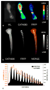
Imaging of bone development in the neonatal mouse in vivo. A d7 mouse was labeled with CatK probe for bone resorption and FRFP750 to denote regions of newly forming bone. A) Left foreleg and paw imaged in white light (WL), CatK680, and FRFP750 demonstrate offset patterns of cathepsin K activation and mineralization in the ulna and radius. Skin present on paw demonstrates significant autofluorescence rather than increased probe activity. B) Tail images showing CatK680, FRFP750, and merged fluorescence. Line in merged imaged refers to C) plot profile of fluorescence of CatK680 and FRFP750, suggesting spatially staggered bone formation and resorption at developing tail vertebral growth plates. Color bars represent fluorescent intensity from low to high.
Figure 4
Figure 4
10 week female Balb/c mice were subjected to OVX, sham, or OVX+daily pamidronate (Pam) injections. MicroCT from proximal tibiae reveals significant loss of bone in OVX animals by day 14. * p<0.05 vs. sham; ** p<0.05 vs. OVX-PAM.
Figure 5
Figure 5
In vivo simultaneous imaging of bone resorption and bone formation in two distinct optical wavelengths. Mice were subjected to OVX and imaged for cathepsin K probe activation 8 or 14 days post OVX. A) White light image of mouse in FMT scanner showing 10 mm tibial region of interest. B) CatK fluorescent signal (yellow) with 10 mm masked tibial region of interest highlighted (rainbow) shows variable intensity at tibial region of interest. C) In 8 day short term bone loss, ovariectomy significantly increases CatK fluorescence over sham, OVX+Pamidronate, and OVX+d-control probe (white bar). Increase in resorption is coupled to increased formation (FRFP750, D). By 14 days post-OVX, resorption has normalized and returned to sham levels (E,F). * p<0.05, ** p<0.001 vs. OVX-CatK probe.
Figure 6
Figure 6
Spatial discrimination of bone resorption and formation in the distal femur. Femora from OVX mice were dissected and imaged via planar fluorescence reflectance imaging (A). Scalebar shows probe fluorescence for FRFP750 and CatK signal scaled to maximum values for each specimen (B) Transverse fluorescence was plotted along the femoral length from the lateral third trochanter (arrow, A) to the distal femoral condyles. Location of peak transverse fluorescence was noted and the distance separating peak cathepsin K and peak FRFP750 signal was found. (C) 8 days post-OVX, cathepsin K probe signal becomes spatially uncoupled from sham and OVX+Pam, and returns to normal by 14 days (D). (E) Schematic representation of uncoupled bone resorption and formation in the distal femur following OVX. In non-OVX conditions, resorption closely follows formation in the growth plate region. Following OVX, the maximal resorption signal (CatK680, red) occurs proximal to the growth plate (arrow) at the site of existing trabeculae. In contrast, the region of maximum formation (FRFP75-, yellow) was not affected by OVX.
Figure 7
Figure 7
Non-decalcified cryosections of trabecular bone within marrow space demonstrates punctate cathepsin K probe staining in the marrow space and adjacent to the trabecular bone (A arrows), while cathepsin K immunostaining reveals punctate staining at comparable locations (B arrows). 20X magnification, scalebar = 50 μm.

Similar articles

Cited by

References

    1. Teitelbaum SL. Bone resorption by osteoclasts. Science. 2000;289:1504–8. - PubMed
    1. Garnero P, Borel O, Byrjalsen I, Ferreras M, Drake FH, McQueney MS, Foged NT, Delmas PD, Delaisse JM. The collagenolytic activity of cathepsin K is unique among mammalian proteinases. Journal of Biological Chemistry. 1998;273:32347–52. - PubMed
    1. Drake FH, Dodds RA, James IE, Connor JR, Debouck C, Richardson S, Lee-Rykaczewski E, Coleman L, Rieman D, Barthlow R, Hastings G, Gowen M. Cathepsin K, but not cathepsins B, L, or S, is abundantly expressed in human osteoclasts. J Biol Chem. 1996;271:12511–6. - PubMed
    1. Gowen M, Lazner F, Dodds R, Kapadia R, Feild J, Tavaria M, Bertoncello I, Drake F, Zavarselk S, Tellis I, Hertzog P, Debouck C, Kola I. Cathepsin K knockout mice develop osteopetrosis due to a deficit in matrix degradation but not demineralization. Journal of Bone & Mineral Research. 1999;14:1654–63. - PubMed
    1. Gelb BD, Shi GP, Chapman HA, Desnick RJ. Pycnodysostosis, a lysosomal disease caused by cathepsin K deficiency. Science. 1996;273:1236–8. - PubMed

Publication types

MeSH terms