. 2009 Jan 30;284(5):3227-3238.
doi: 10.1074/jbc.M806177200. Epub 2008 Nov 13.

Calcium-dependent binding of HCN1 channel protein to hair cell stereociliary tip link protein protocadherin 15 CD3

Affiliations

Calcium-dependent binding of HCN1 channel protein to hair cell stereociliary tip link protein protocadherin 15 CD3

Neeliyath A Ramakrishnan et al. J Biol Chem. .

Abstract

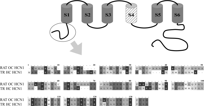
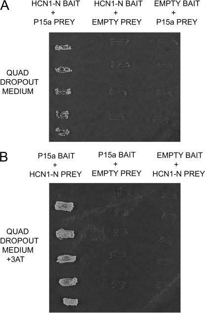
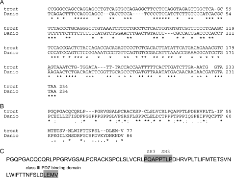
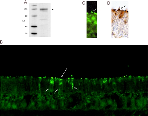
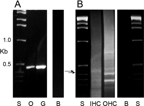
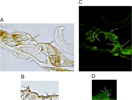
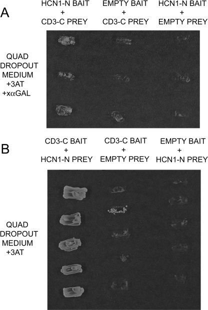
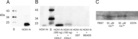
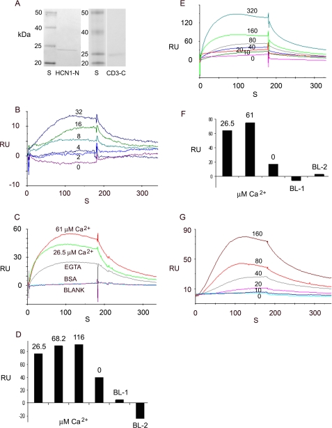
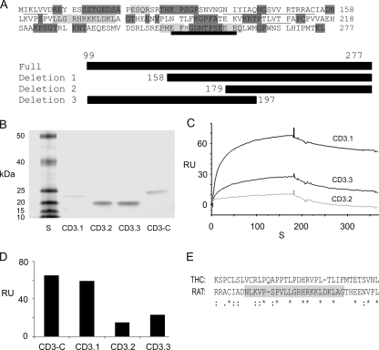
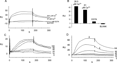
The cytoplasmic amino terminus of HCN1, the primary full-length HCN isoform expressed in trout saccular hair cells, was found by yeast two-hybrid protocols to bind the cytoplasmic carboxyl-terminal domain of a protocadherin 15a-like protein. HCN1 was immunolocalized to discrete sites on saccular hair cell stereocilia, consistent with gradated distribution expected for tip link sites of protocadherin 15a. HCN1 message was also detected in cDNA libraries of rat cochlear inner and outer hair cells, and HCN1 protein was immunolocalized to cochlear hair cell stereocilia. As predicted by the trout hair cell model, the amino terminus of rat organ of Corti HCN1 was found by yeast two-hybrid analysis to bind the carboxyl terminus of protocadherin 15 CD3, a tip link protein implicated in mechanosensory transduction. Specific binding between HCN1 and protocadherin 15 CD3 was confirmed with pull-down assays and surface plasmon resonance analysis, both predicting dependence on Ca(2+). In the presence of calcium chelators, binding between HCN1 and protocadherin 15 CD3 was characterized by a K(D) = 2.39 x 10(-7) m. Ca(2+) at 26.5-68.0 microm promoted binding, with K(D) = 5.26 x 10(-8) m (at 61 microm Ca(2+)). Binding by deletion mutants of protocadherin 15 CD3 pointed to amino acids 158-179 (GenBank accession number XP_238200), with homology to the comparable region in trout hair cell protocadherin 15a-like protein, as necessary for binding to HCN1. Amino terminus binding of HCN1 to HCN1, hypothesized to underlie HCN1 channel formation, was also found to be Ca(2+)-dependent, although the binding was skewed toward a lower effective maximum [Ca(2+)] than for the HCN1 interaction with protocadherin 15 CD3. Competition may therefore exist in vivo between the two binding sites for HCN1, with binding of HCN1 to protocadherin 15 CD3 favored between 26.5 and 68 microm Ca(2+). Taken together, the evidence supports a role for HCN1 in mechanosensory transduction of inner ear hair cells.


Figures

FIGURE 1.
FIGURE 1.
Alignment (with OMIGA 2.0) of the amino terminus of rat organ of Corti HCN1 and trout saccular hair cell HCN1 (GenBank™ accession number AAQ04053), both used as bait in yeast two-hybrid protocols, pull-down assays and SPR analyses. The cytoplasmic amino terminus sequence is encircled in a schematic depiction of HCN1 with S1-S6 transmembrane regions and S4 (hatched) representing the voltage sensor. For rat organ of Corti HCN1, aa 11-18 display EF hand-like characteristics. Amino acids 11 and 17 are designated as having low α-helical character, whereas aa 12-16 have loop character (PROF predictions (47)). SRD (aa 13-15) and DGN (aa 16-18) have been cited as Ca2+-binding motifs (48).
FIGURE 2.
FIGURE 2.
Interaction of a protocadherin 15a-like sequence with trout saccular hair cell HCN1 amino-terminal cytoplasmic domain. A, first lane, binding of aa 1-118 of HCN1-N as bait to a protocadherin 15a-like protein as prey, determined on quadruple drop-out medium with yeast two-hybrid mating protocols. Second lane, negative control with HCN1-N as bait and empty prey vector. Third lane, negative control with empty bait vector paired with prey sequence. B, reversal of bait and prey. First lane, protocadherin 15a-like protein as bait and HCN1-N as prey, on high stringency quadruple drop-out medium (with 2 mm 3-AT). Second lane, negative control with protocadherin 15a as bait and empty prey vector. Third lane, negative control in which empty bait vector is paired with the HCN1-N prey sequence.
FIGURE 3.
FIGURE 3.
Nucleotide and amino acid sequences of the trout saccular hair cell protocadherin 15a-like protein compared with corresponding sequences of D. rerio (GenBank™ accession number AY772390). A, comparison of nucleotide sequences of the carboxyl terminus of protocadherin 15a aligned with Clustal. B, comparison of amino acid sequences of the carboxyl terminus of protocadherin 15a for trout and D. rerio. C, indication of two SH3 binding sites, PQAP, PTLP, and PDZ3-interacting sequence LEMV in the trout saccular hair cell aa sequence.
FIGURE 4.
FIGURE 4.
Immunodetection of HCN1 protein in the trout saccular hair cells. A, Western blot of HCN1 protein in trout saccular hair cell layer. A custom rabbit polyclonal antibody (1:1,250), targeting a 19-aa peptide found in the carboxyl terminus of saccular hair cell HCN1, immunolabeled a protein of predicted molecular mass (104 kDa, arrow), based upon full-length HCN1 sequence (1) (GenBank™ accession number AF421883). B, immunofluorescence localization of HCN1 in the trout saccular sensory epithelium. Immunoreactivity for HCN1 was found in saccular hair cells at the apical membrane (long arrow) extending along the lateral cell membranes to basal sites of the hair cells (short arrows), the latter consistent in position to sites opposing efferent innervation. Sometimes, circular patterns were observed when the plane of sectioning of the epithelium was tilted, possibly corresponding to the edges of the cuticular plate. C, HCN1 was immunolocalized with immunofluorescence to the hair cell stereocilia (arrow), with foci of immunofluorescence present at gradated sites on the stereociliary array. D, immunoreactivity for HCN1 was also detected with 3,3′-diaminobenzidine, with discrete puncta present in stereociliary arrays (arrow).
FIGURE 5.
FIGURE 5.
HCN1 message expressed in rat organ of Corti and cochlear hair cells. A, amplification with PCR of a 455-bp cDNA from microdissected organ of Corti (lane O) and spiral ganglion (lane G) subfractions from the rat cochlea, with 1-kb standards (lane S) and negative control in which cDNA was omitted (lane B). B, HCN1 message in cochlear inner hair cells and outer hair cells. HCN1 message in λ-ZAP cDNA libraries of rat cochlear inner hair cells (lane IHC) and outer hair cells (lane OHC). The outer hair cell ∼420-bp amplification product (lower arrow) and inner hair cell 455-bp amplification product (upper arrow), the latter with application of nested primers, yielded rat HCN1 sequence. Lane S, standard; lane B, water blank.
FIGURE 6.
FIGURE 6.
HCN1 immunoreactivity within the organ of Corti of the rat cochlea. A, HCN1 is immunolocalized to nerve fibers approaching and passing the habenula perforata (long arrow) en route to the base of the inner hair cell (intermediate length arrow). Immunoreactivity was also localized to stereocilia of the inner hair cell (short arrow) and outer hair cell (short arrow + asterisk). B, HCN1 immunoreactivity is associated with stereocilia of outer hair cells (short arrow), as well as inner hair cells (intermediate length arrow). C, HCN1-immunoreactive nerve fibers approach and cross the habenula perforata (long arrow) and follow pathways to sites close to the base of the inner hair cell (intermediate length arrow). Immunoreactivity is observed at the base of outer hair cells overlapping Deiters' cells (short arrows) and is associated with the inner hair cell stereociliary array (short arrow + asterisk; green fluorescence). Yellow fluorescence overlapping apical sites of the inner hair cell represents autofluorescence, likely from flavin adenine dinucleotide (49). D, foci of HCN1 immunoreactivity (arrows) associated with the stereociliary array of an inner hair cell.
FIGURE 7.
FIGURE 7.
HCN1 amino-terminal cytoplasmic domain interacts with protocadherin 15 CD3 carboxyl-terminal domain in yeast two-hybrid assay. A, rat organ of Corti HCN1-N as bait and rat organ of Corti CD3-C as prey in selection medium SD/-Trp/-Leu/-His/-Ade containing 2 mm 3-AT and α-galactoside. The first lane shows expression of the Gal-4 marker, which is expressed only when there is interaction between the bait and prey fusion peptides. The second and third lanes are negative controls in which the bait and prey sequences, respectively, are replaced by empty vector. B, reversal of bait and prey. First lane, CD3-C as bait and HCN1-N as prey in selection medium Sd/-Trp/-Leu/-His/-Ade containing 2 mm 3-AT. The second and third lanes are negative controls, CD3-C as bait and empty prey vector and HCN1-N as prey and empty bait vector, respectively.
FIGURE 8.
FIGURE 8.
GST pulldown assay of binding between the amino terminus of HCN1 and the carboxyl terminus of protocadherin 15CD3, both produced from rat organ of Corti cDNA. A, Western blot detection of the amino terminus fusion protein. First lane, standards of 23, 34, and 43 kDa (Santa Cruz). Second lane, prominent band identifies amino terminus of HCN1 fusion protein, predicted mass of 23 kDa, detected with affinity-purified rabbit polyclonal antibody at 1:200 (APC-056, Alomone Labs, Israel), targeting amino acids 6-24 of rat HCN1. The predicted mass of 23 kDa arises from 19 kDa HCN1 +∼4-kDa vector protein. B, HCN1-N pulldown with GST-protocadherin 15 CD3-C fusion protein in PBST. First lane, purified HCN1-N fusion product used in the pull-down assay, detected with an antipolyhistidine monoclonal antibody. Second lane (S), protein standards, molecular masses 23, 34, and 43 kDa. Third lane, binding of HCN1-N fusion protein (∼350 ng) with GST-protocadherin 15 CD3-C cleared lysate, pulled down with glutathione-Sepharose, and detected with antipolyhistidine monoclonal antibody. Fourth lane, binding of HCN1-N fusion protein (∼100 ng) with GST-protocadherin 15 CD3-C lysate. Fifth lane, HCN1-N (∼350 ng) incubated with GST bacterial lysate as a negative control. Sixth lane, rat HCN1-N fusion protein mixed with Sepharose beads under the same conditions as above, representing another negative control. C, HCN1-N pulldown with CD3-C in PBST, 53 μm Ca2+, 93 μm Ca2+, or EGTA. 200 μl of HCN1-N lysate (corresponding to ∼0.8 mg of total bacterial protein) and 250 μl of GST-CD3-C lysate (representing ∼0.8 mg of total bacterial protein) were used for each pull-down reaction.
FIGURE 9.
FIGURE 9.
SPR analysis of binding interaction between HCN1 and protocadherin 15 CD3. A, affinity-purified fusion proteins. First lane, prestained protein standards on SDS-PAGE gel: 20, 25, 40, and 50 kDa (Bench-Mark, Invitrogen). Second lane, affinity-purified rat organ of Corti HCN1-N fusion protein at 23 kDa, stained with Coomassie Blue. Third lane, protein standards on SDS-PAGE gel. Fourth lane, affinity-purified rat organ of Corti protocadherin 15 CD3-C fusion protein at 24 kDa, stained with Coomassie Blue. B, binding between the amino terminus of rat organ of Corti HCN1 (HCN1-N) as ligand and the carboxyl terminus of rat organ of Corti protocadherin 15CD3 (CD3-C) as analyte as a function of CD3-C concentration in the presence of 3 mm EDTA. For sensorgram, the response units (RU) are indicated for 0 (violet), 2 (blue), 4 (gray-blue), 8 (turquoise), 16 (green), and 32 (purple) nm analyte. KD = 3.57 × 10⁻⁷ m. C, the binding of HCN1-N (ligand) to CD3-C (analyte) (50 nm) has aCa2+-dependent component. Response units are indicated for binding with CD3-C at 61 μm Ca2+ (red), 26.5 μm Ca2+ (green), or 1 mm EGTA (gray), compared with the response for bovine serum albumin (turquoise) or blank buffer (magenta). D, Ca2+ dependence of the response (RU) for interaction of CD3-C (analyte) with HCN1-N (ligand). The bar graph indicates the response for 150 nm CD3-C at 26.5 μm Ca2+, 68.2 μm Ca2+, 116 μm Ca2+, and 1 mm EGTA, compared with blank buffer (BL-1, BL-2). E, kinetics of binding between HCN1-N (ligand) and CD3-C (analyte) at 61 μm Ca2+:0 (pink), 10 (red), 20 (gray-green), 40 (blue), 80 (gray), 160 (green), and 320 (turquoise) nm analyte. KD = 2.3 × 10⁻⁸ m. F, bar graph depicting response with reversal of ligand and analyte. Ca2+-dependent binding of CD3-C (ligand) and HCN1-N (analyte) (100 nm) at 26.5 μm Ca2+, 61 μm Ca2+, and 1 mm EGTA, compared with responses for buffer with 1 mm EGTA + 1 mm Ca2+ (BL-1) or buffer alone (BL-2). G, kinetic series for binding between CD3-C (ligand) and HCN1-N (analyte) at 61 μm Ca2+ for 0 (turquoise), 10 (blue), 20 (pink), 40 (gray), 80 (red), and 160 (brown) nm analyte. KD = 3.41 × 10⁻⁸ m.
FIGURE 10.
FIGURE 10.
Deletion mutants of rat organ of Corti protocadherin CD3 implicate a 22-amino acid peptide within the carboxyl terminus as a requirement for binding. A, amino acid sequence for the carboxyl terminus segment of protocadherin 15 CD3 used as prey. The secondary structure is indicated: underlining, β-sheets; light gray shading, α-helix; dark gray shading, loop; thick underlining, aa 248-259 comprising a putative EF hand Ca2+-binding motif (50). There was 11% helix, 19% β-strand, and 70% loop sequence. The full-length sequence (CD3-C) and deletion mutants 1 (CD3.1), 2 (CD3.2), and 3 (CD3.3) expressed in pRSET-A vector are indicated by heavy black bars. B, Coomassie Blue staining of SDS-PAGE gel of affinity-purified mutants. S, standards: 10, 15, 20, 25, 40, and 50 kDa (Invitrogen). The molecular mass in kDa is indicated for CD3.1, CD3.2, CD3.3, and CD3-C. C, binding of deletion mutants of the protocadherin 15 CD3-C (CD3.1, CD3.3, and CD3.2, all as analytes at 120 nm) to HCN1-N at 26.5 μm Ca2+. D, comparison of SPR responses for CD3-C, CD3.1, CD3.2, and CD3.3. Overall, CD3-C versus CD3.1, 0.05 < p < 0.10, n = 5; CD3.1 versus CD3.2, p < 0.05, n = 7; CD3.1 versus CD3.3, 0.05 < p < 0.10, n = 7; CD3.3 versus CD3.2, p = 0.103, n = 6 (two-tailed paired-variate analysis). E, clustal alignment of amino acids in rat protocadherin 15CD3-C and trout protocadherin 15a. The 22-aa peptide region in the carboxyl terminus of rat protocadherin 15 CD3-C (highlighted in gray), implicated as important for binding to HCN1-N, has homology to protocadherin 15a-like protein expressed in trout saccular hair cells.
FIGURE 11.
FIGURE 11.
Rat organ of Corti HCN1 binds to itself via its amino terminus, and the binding is Ca2+-dependent. A, SPR sensorgram for HCN1-N (200 nm) as analyte, binding to HCN1-N as ligand, at 26.5 μm Ca2+, 61 μm Ca2+, and with EGTA (1 mm), compared with the response with buffer alone (26.5 μm Ca2+). B, bar graph representation in same order as for A. C, kinetic series of binding interactions for HCN1-N as analyte to HCN1-N as ligand (at 61 μm Ca2+) for 0, 10, 20, 40, 80, and 160 nm analyte. D, comparable kinetic series of binding interactions for CD3-C as analyte with HCN1-N as ligand (at 61 μm Ca2+) in a back-to-back comparison to C for 0, 10, 20, 40, 80, and 160 nm analyte.

Similar articles

Cited by

References

    1. Cho, W. J., Drescher, M. J., Hatfield, J. S., Bessert, D. A., Skoff, R. P., and Drescher, D. G. (2003) Neuroscience 118 525-534 - PubMed
    1. Holt, J. R., and Eatock, R. A. (1995) J. Neurophysiol. 73 1484-1502 - PubMed
    1. Farris, H. E., Wells, G. B., and Ricci A. J. (2006) J. Neurosci. 26 12526-12536 - PMC - PubMed
    1. Ramakrishnan, N. A., Drescher, M. J., and Drescher, D. G. (2007) Assoc. Res. Otolaryngol. Abstr. 30 31
    1. Proenza, C., Angoli, D., Agranovich, E., Macri, V., and Accili, E. A. (2002) J. Biol. Chem. 277 5101-5109 - PubMed

Publication types

MeSH terms