. 2009 Mar;150(3):1202-16.
doi: 10.1210/en.2008-0293. Epub 2008 Nov 13.

Orexin-A hyperphagia: hindbrain participation in consummatory feeding responses

Affiliations

Orexin-A hyperphagia: hindbrain participation in consummatory feeding responses

John-Paul Baird et al. Endocrinology. 2009 Mar.

Abstract

Orexin-A (ORXA) is an orexigenic neuropeptide produced by the lateral hypothalamus that increases food intake when injected into the brain ventricles or forebrain nuclei. We used a licking microstructure analysis to evaluate hindbrain and forebrain ORXA effects in intact and hindbrain-lesioned rats, to identify the motivational and anatomical bases of ORXA hyperphagia. Intact rats with cannulas in the fourth brain ventricle (4V) received vehicle (artificial cerebrospinal fluid) or ORXA (0.1, 0.4, 1, or 10 nm) injections before 90 min access to 0.1 m sucrose. Meal size and frequency were increased in a double-dissociated manner by the 1 and 10 nm doses, respectively. In experiment 2, 4V 1 nm ORXA was applied to rats offered solutions varied in caloric and gustatory intensity (water and 0.1 and 1 m sucrose). ORXA increased meal frequency for all tastants. ORXA increased meal size only for 0.1 m sucrose, by prolonging the meal without affecting early ingestion rate or lick burst size, suggesting that 4V ORXA influenced inhibitory postingestive feedback rather than taste evaluation. In experiment 3, rats with cannulas in the third ventricle (3V) received dorsal medullary lesions centered on the area postrema (APX group) or sham procedures, and licking for water and 0.1 and 1 m sucrose was evaluated after 1 nm 3V ORXA/artificial cerebrospinal fluid injections. The 3V ORXA increased 0.1 m sucrose meal size and meal frequency for all tastants in the sham group, as observed after 4V ORXA in experiment 2. In the APX group, 3V ORXA injections influenced meal frequency, but they no longer increased meal size. However, the APX rats increased meal size for 0.1 m sucrose after food and water deprivation and after 3V angiotensin II injection. They also showed meal size suppression after 3V injection of the melanocortin-3/4 receptor agonist melanotan II (1 nm). These findings suggest that the area postrema and subjacent nucleus of the solitary tract are necessary for increases in consummatory (meal size) but not appetitive (meal frequency) responses to 3V ORXA. The meal size increases may be due to reduced postingestive feedback inhibition induced by ORXA delivered to either the hindbrain or forebrain ventricles.


Figures
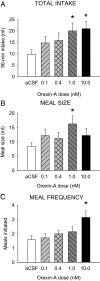
Figure 1
Figure 1
ORXA infused to the 4V increased the consumption of 0.1 m sucrose during the test session to a similar extent at the 1 and 10 nm doses, but this result was achieved through separate underlying dose-dependent processes. A, Mean (+ se) 90-min intake (milliliters) values for aCSF (white bar) and four doses of ORXA (hatched and black bars) in 4V cannula-fitted rats (n = 7) ingesting 0.1 m sucrose solution. *, P < 0.05. B, Mean (+se) meal size (milliliters) for the same conditions as in A. *, P < 0.05. C, Mean (+se) number of meals initiated for the test solution for the same conditions as in A. *, P < 0.05.
Figure 2
Figure 2
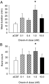
The increase in meal size induced by 4V 1 nm ORXA was achieved through increases in the meal duration and the number of licking bursts in the meal. A, Mean (+se) meal duration (minutes) values for aCSF (white bar) and four doses of ORXA (hatched and black bars) in 4V cannula-fitted rats (n = 7) ingesting 0.1 m sucrose solution [ANOVA: F(4,24) = 2.99, P < 0.04; aCSF-1 nm comparison: *, P < 0.05]. B, Mean (+se) number of bursts in the meal for the same conditions as in A [ANOVA: F(4,24) = 1.09, NS; aCSF-1 nm comparison: *, P < 0.04].
Figure 3
Figure 3
ORXA (1 nm) infused to the 4V differentially increased the meal size or meal frequency in a concentration-dependent manner. A, Mean (+ se) meal size (milliliters) values after aCSF (white bar) and ORXA (black bars) injections in 4V cannula-fitted rats (n = 16) ingesting water and 0.1 and 1 m sucrose solutions [ANOVA: concentration F(2,30) = 26.49, P < 0.001; drug F(1,15) = 8.74, P < 0.01; interaction F(2,30) = 5.26, P < 0.01; 0.1 m pairwise comparison: t(15) = −3.22; *, P = 0.006]. B, The mean (+se) number of meals initiated in the test session for the same conditions as in A did not vary by tastant concentration [F(2,30) = 0.12, NS], but it was increased for all tastants after ORXA [drug F(1,15) = 43.95; *, P < 0.001; interaction F(2,30) = 1.25, NS].
Figure 4
Figure 4
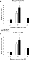
ORXA (1 nm) infused to the 4V increased 0.1 m sucrose meal consumption through modification of measures associated with postingestive feedback inhibition. A, Mean (+se) meal duration (minutes) after aCSF (white bar) and ORXA (black bars) injections in 4V cannula-fitted rats (n = 16) ingesting water and 0.1 and 1 m sucrose solutions (ANOVA: concentration F(2,30) = 15.52, P < 0.001; drug F(1,15) = 11.22, P < 0.004; interaction F(2,30) = 3.33; *, P < 0.05. B, Mean (+ se) number of licking bursts for the same conditions as in A, ORXA significantly increased the number of bursts, primarily for 0.1 m sucrose (concentration F(2,30) = 10.24, P < 0.001; drug F(1,15) = 8.89, P = 0.009; interaction F(2,30) = 2.84, P = 0.07; 0.1 m sucrose comparison t(15) = −2.34; *, P = 0.03).
Figure 5
Figure 5
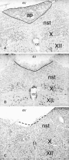
Example photomicrographs depicting the range of lesions observed (×40 magnification). A, A brain from a sham lesioned animal reveals intact AP, NST, DMX (X), and hypoglossal motor nucleus (XII). CC, Central canal. B, Image (same level as A) from the brain of a rat in the lesion group typical of those exhibiting the most minimal damage. Here, the AP was considerably but incompletely destroyed. C, Image (same level as A) from the brain of a rat in the lesion group typical of those exhibiting the most extensive damage. In this animal, the AP was completely ablated, with collateral damage to the subjacent dorsal medial and commissural subnuclei of the NST, particularly on the left side. The border of the AP/NST is suggested by the dashed line.
Figure 6
Figure 6
AP lesions abolished the hyperphagic response to 3V injection of ORXA (1 nm). A, Mean (+ se) meal size (milliliters) values after aCSF (white bar) and ORXA (black bars) injections in 3V cannula-fitted sham operated rats (n = 8) ingesting water and 0.1 and 1 m sucrose solutions. ORXA increased meal size overall [drug F(1,7) = 16.91; P < 0.005] and specifically for 0.1 m sucrose [t(7) = −2.61; *, P = 0.035]. B, Same conditions as A for 3V cannula-fitted AP/NST lesioned rats (APX; n = 17). There was no significant effect of drug [F(1,16) = 0.80, NS] and no significant interaction [F(2,32) = 0.70, NS], and the paired comparison for 0.1 m sucrose was not significant [t(16) = −0.86, NS].
Figure 7
Figure 7
The increase in 0.1 m sucrose meal duration after 3V infusion of ORXA (1 nm) in sham operated rats was attenuated in rats with AP/NST lesions (APX). A, Mean (+ se) meal duration (minutes) values after aCSF (white bars) and ORXA (black bars) injections in 3V cannula-fitted sham operated rats (n = 8) ingesting water and 0.1 and 1 m sucrose solutions. Meal duration was specifically increased by ORXA for 0.1 m sucrose [ANOVA: concentration F(2,14) = 21.90, P < 0.001; drug F(1,7) = 4.70, P = 0.067; interaction F(2,14) = 7.14, P = 0.007; 0.1 m sucrose paired comparison t(7) = −2.52; *, P = 0.04]. B, Same conditions as A for 3V cannula-fitted APX rats (n = 17) [ANOVA: concentration F(2,32) = 9.78, P < 0.001; drug F(1,16) = 8.26, P = 0.01; interaction [F(2,32) = 1.20, NS; 0.1 m sucrose paired comparison t(7) = −1.67, NS].
Figure 8
Figure 8
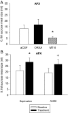
A, Infusion of MTII (1 nm) significantly reduced meal size in AP/NST lesioned (APX) rats [t(9) = 2.46; P < 0.04], but ORXA (1 nm) infusions were without effect [t(9) = −0.85, NS]. Mean (+ se) meal size for the MTII-tested subgroup (n = 10) is shown. B, APX rats did increase mean (+ se) meal size for hypotonic 0.1 m sucrose after 22.5h food and fluid deprivation [t(9) = −2.75; *, P = 0.023], and they exhibited a marginally significant trend in the same direction after angiotensin II [ANGII; t(9) = −2.21; +, P = 0.055].

Similar articles

Cited by

References

    1. Kunii K, Yamanaka A, Nambu T, Matsuzaki I, Goto K, Sakurai T 1999 Orexins/hypocretins regulate drinking behaviour. Brain Res 842:256–261 - PubMed
    1. Krowicki ZK, Burmeister MA, Berthoud HR, Scullion RT, Fuchs K, Hornby PJ 2002 Orexins in rat dorsal motor nucleus of the vagus potently stimulate gastric motor function. Am J Physiol Gastrointest Liver Physiol 283:G465–G472 - PubMed
    1. Miyasaka K, Masuda M, Kanai S, Sato N, Kurosawa M, Funakoshi A 2002 Central orexin-A stimulates pancreatic exocrine secretion via the vagus. Pancreas 25:400–404 - PubMed
    1. Rodgers RJ, Ishii Y, Halford JC, Blundell JE 2002 Orexins and appetite regulation. Neuropeptides 36:303–325 - PubMed
    1. Sakurai T, Amemiya A, Ishii M, Matsuzaki I, Chemelli RM, Tanaka H, Williams SC, Richarson JA, Kozlowski GP, Wilson S, Arch JR, Buckingham RE, Haynes AC, Carr SA, Annan RS, McNulty DE, Liu WS, Terrett JA, Elshourbagy NA, Bergsma DJ, Yanagisawa M 1998 Orexins and orexin receptors: a family of hypothalamic neuropeptides and G protein-coupled receptors that regulate feeding behavior. Cell 92:1, page following 696 - PubMed

Publication types

MeSH terms