. 2008 Nov 18;105(46):17818-23.
doi: 10.1073/pnas.0804446105. Epub 2008 Nov 13.

The G-protein-coupled receptor kinase 5 inhibits NFkappaB transcriptional activity by inducing nuclear accumulation of IkappaB alpha

Affiliations

The G-protein-coupled receptor kinase 5 inhibits NFkappaB transcriptional activity by inducing nuclear accumulation of IkappaB alpha

Daniela Sorriento et al. Proc Natl Acad Sci U S A. .

Abstract

G-protein-coupled receptor (GPCR) kinases, GRKs, are known as serine/threonine kinases that regulate GPCR signaling, but recent findings propose functions for these kinases besides receptor desensitization. Indeed, GRK5 can translocate to the nucleus by means of a nuclear localization sequence, suggesting that this kinase regulates transcription events in the nucleus. To evaluate the effect of GRK5-IkappaB alpha interaction on NFkappaB signaling, we induced the overexpression and the knockdown of GRK5 in cell cultures. GRK5 overexpression causes nuclear accumulation of IkappaB alpha, leading to the inhibition of NFkappaB transcriptional activity. Opposite results are achieved by GRK5 knockdown through siRNA. A physical interaction between GRK5 and IkappaB alpha, rather than phosphorylative events, appears as the underlying mechanism. We identify the regulator of gene protein signaling homology domain of GRK5 (RH) and the N-terminal domain of IkappaB alpha as the regions involved in such interaction. To confirm the biological relevance of this mechanism of regulation for NFkappaB, we evaluated the effects of GRK5-RH on NFkappaB-dependent phenotypes. In particular, GRK5-RH overexpression impairs apoptosis protection and cytokine production in vitro and inflammation and tissue regeneration in vivo. Our results reveal an unexpected role for GRK5 in the regulation of NFkappaB transcription activity. Placing these findings in perspective, this mechanism may represent a therapeutic target for all those conditions involving excessive NFkappaB activity.


Conflict of interest statement

The authors declare no conflict of interest.

Figures

Fig. 1.
Fig. 1.
GRK5 binds and stabilizes IκBα, causing inhibition of NFκB activity. (A) IκBα and GRK5 levels were analyzed in whole, cytosolic, and nuclear extracts by Western blot in BAEC overexpressing GRK5-WT. The overexpression of GRK5-WT increases IκBα levels in whole-cell extracts and induces IκBα nuclear accumulation. (B) To evaluate the effects of GRK5 on IκBα turnover, we analyzed GRK5 and IκBα expression in a time-course of GRK5 transfection. The increase of GRK5 levels induces a progressive increase of IκBα levels and a reduction of NFκB activity (*P < 0.05 vs. control). (C) A time course of transfection of GRK5 was performed in BAEC and GRK5 and IκBα levels analyzed both in whole- and nuclear-cell extracts. The time-dependent increase of GRK5 levels associates to IκBα stabilization and nuclear localization and subsequently NFκB activity inhibition (*P < 0.05 vs. control). (D) HEK293 transfected with GRK5-WT or GRK5siRNA were analyzed by WB for GRK5 and IκBα expression. As in BAEC, GRK5 overexpression induces IκBα accumulation; conversely, GRK5 knockdown by GRK5siRNA associates to IκBα degradation. Actin is shown for protein-loading control. HEK293 were transfected with κB-Luc plasmid and GRK5-WT or GRK5-siRNA and luciferase activity was measured. GRK5-WT overexpression causes inhibition of NFκB activity, whereas GRK5 knockdown increases NFκB transcription levels. The data in the bar graph are expressed as mean±SEM and are representative of 3 experiments (*P < 0.05 vs. control). (E) GRK5 and IκBα expression and luciferase activity were assessed in HEK293 transfected with increasing doses of GRK5siRNA. The progressive knockdown of GRK5 associates to a similar reduction in IκBα levels and NFκB activity. Actin was used as loading control (*P < 0.05 vs. control). (F) BAEC were transiently transfected with either GRK5-WT or the kinase-dead mutant GRK5-K215R. Both variants are found in the nucleus together with increased amount of IκBα and NFκB. Equal amounts of proteins are confirmed by WB for histone 3. BAEC were transfected with κB-Luc plasmid and GRK5-WT, GRK5-K215R, and GRK2, and luciferase activity was measured. Overexpression of both GRK5-WT or GRK5-K215R significantly inhibit NFκB transcription activity in BAEC, whereas GRK2 overexpression has no inhibitory effect. The data are expressed as mean±SEM and are representative of three experiments (*P < 0.05 vs. control).
Fig. 2.
Fig. 2.
The RH domain of GRK5 interacts with IκBα and causes inhibition of NFκB activity. (A) To map the IκBα binding region on GRK5, we created myc/histidine-tagged, truncated mutants of GRK5 including GRK5-WT amino acids 1–590, GRK5-NT amino acids 1–176, GRK5-RH amino acids 50–176, and GRK5-CT amino acids 176–590. To verify the hypothesis that GRK5 masks the N-NES on IκBα, we created myc/histidine-tagged mutants of IκBα including IκBα-WT amino acids 1–317 and IκBα-CT amino acids 59–317. (B) In lysates from BAEC overexpressing histidine-tagged GRK5-WT, -NT, -RH, and -CT, histidine was precipitated by using Ni Sepharose beads and subjected to a Western blot with anti-IκBα or anti-His antibodies. GRK5-WT, -NT, and -RH (but not -CT) coprecipitate with IκBα. (C) Whole extracts from control and GRK5-RH transfected cells were immunoprecipitated with anti-IκBα antibody, and Western blot was performed with anti-GRK5 antibody. GRK5-RH overexpression attenuates IκBα immunoprecipitation of GRK5 (*P < 0.05 vs. control). (D) The immunoprecipitation study with IκBα mutants shows that IκBα-WT coimmunoprecipitates with GRK5 whereas IκBα-CT loses such ability. Both IκBα-WT and its mutant preserve the ability to interact with NFκB. (E) We evaluated the effects of GRK5 on NFκB activity by luciferase assay in BAEC overexpressing truncated GRK5 mutants and stimulated with LPS (1 μg/ml) for 4 h. GRK5-WT, -NT, and -RH inhibit LPS-induced NFκB transcriptional activity whereas GRK5-CT doesn't change this response (*P < 0.05). (F) To further confirm such an effect, we evaluated LPS-induced NFκB binding to DNA by electrophoretic mobility shift assay in nuclear extracts. GRK5-WT, -NT, and -RH (but not -CT) overexpression cause a significant inhibition of LPS-induced NFκB DNA binding. Data are expressed as mean±SEM (*P < 0.05 vs. control).
Fig. 3.
Fig. 3.
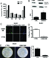
GRK5-RH regulates different phenotypes that are under the transcriptional control of NFκB. (A) NFκB-dependent TNFα expression was assessed by Northern blot in BAEC stimulated with LPS for 4 h. GRK5-WT, -NT, and -RH (but not -CT) inhibit TNFα mRNA expression induced by LPS (*P < 0.05). (B) To evaluate the effect of GRK5-RH on apoptosis, we analyzed the cleavage of caspase 3 by Western blot. The overexpression of GRK5-RH increases cleaved caspase 3 levels, suggesting that NFκB inhibition induced by GRK5-RH causes an increase of apoptotic responses (*P < 0.05 vs. control (C) GRK5-RH overexpression causes apoptosis as shown by Annexin-V staining respective to live cells; propidium iodide and DAPI were used as controls. (D) Migration of confluent BAEC was measured after the cell monolayer was partially wiped away. The area of the migrating cells was measured in several fields of view and is shown in the graph. Data are presented as FBS-induced percentages of migration respective to resting cells (*P < 0.05 vs. FBS 5%). (E) Representative-phase contrast photomicrographs of control and GRK5-RH transiently transfected BAEC plated on Matrigel are shown. Microscopy revealed numbers of network projections formed in each group after 12 h of incubation (*P < 0.05 vs. control). Data from three experiments in triplicate are summarized in the graph.
Fig. 4.
Fig. 4.
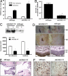
AdGRK5-NT inhibits regenerative responses in vivo. (A) After surgical removal of the femoral artery in rats, blood flow through the ischemic hindlimb is granted by neoangiogenic and vascular regenerative phenomena, mediated in part by NFκB-dependent cytokines. After 14 days of chronic ischemia, the time to perfusion of the ischemic hindlimb was assessed by digital angiographies, showing an increased TIMI score to perfusion in AdGRK5-NT respective to AdEmpty rats (*P < 0.05). (B) Blood flow was also assessed by muscle content of dyed beads after intra-arterial injection in the bloodstream. AdGRK5-NT reduces blood perfusion in ischemic hindlimb respective to AdEmpty. The ischemic-to-nonischemic ratio of dyed beads content per mg of hindlimb muscle tissue (*P < 0.05 vs. AdEmpty) is shown. (C) RNA from the hindlimbs of AdEmpty and AdGRK5-NT-treated rats was extracted by means of trizol reagent and TNFα expression was evaluated by Northern blot. The analysis shows an attenuation of cytokine expression in GRK5-NT-treated ischemic compared to AdEmpty ischemic hindlimb (*P < 0.05). (D) In another model of regeneration, the skin-wound healing, we observed a longer period for healing of wounds that are treated with AdGRK5-NT rather than AdEmpty at the time of surgery. The expression of the transgene was assessed by taking advantage of the bicistronic nature of the adenovirus, encoding also for the GFP under the CMV promoter, with the specific antibody anti-GFP. The transgene localizes both in epidermis and dermis. (E) In AdGRK5-NT-treated wounds, haematoxylin/eosin staining reveals a smaller amount of infiltrate when compared with AdEmpty control. (F) Similarly, MCP-1 expression assessed in the wound is reduced by AdGRK5-NT when compared with AdEmpty wounds.

References

    1. Birbach A, et al. Signaling molecules of the NF-kappa B pathway shuttle constitutively between cytoplasm and nucleus. J Biol Chem. 2002;277:10842–10851. - PubMed
    1. Claing A, Laporte SA, Caron MG, Lefkowitz RJ. Endocytosis of G protein-coupled receptors: roles of G protein-coupled receptor kinases and beta-arrestin proteins. Prog Neurobiol. 2002;66:61–79. - PubMed
    1. Ghosh S, Karin M. Missing pieces in the NF-kappaB puzzle. Cell. 2002;109(Suppl):S81–S96. - PubMed
    1. Pitcher JA, Freedman NJ, Lefkowitz RJ. G protein-coupled receptor kinases. Annu Rev Biochem. 1998;67:653–692. - PubMed
    1. Penn RB, Pronin AN, Benovic JL. Regulation of G protein-coupled receptor kinases. Trends Cardiovasc Med. 2000;10:81–89. - PubMed

Publication types

MeSH terms