. 2008 Dec 17;27(24):3311-21.
doi: 10.1038/emboj.2008.241. Epub 2008 Nov 13.

Interferon-inducible protein, P56, inhibits HPV DNA replication by binding to the viral protein E1

Affiliations

Interferon-inducible protein, P56, inhibits HPV DNA replication by binding to the viral protein E1

Fulvia Terenzi et al. EMBO J. .

Abstract

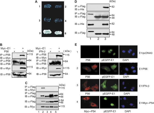
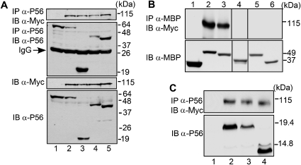
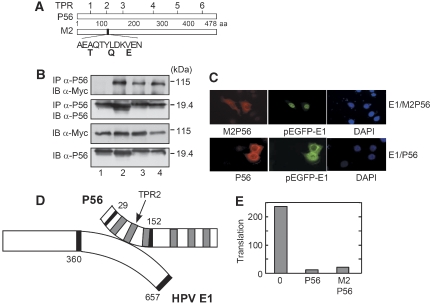
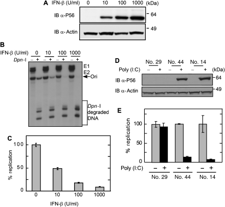
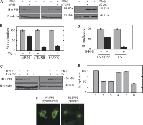
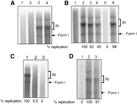
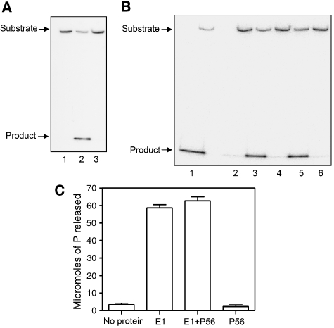
Type I interferon (IFN) inhibits, by an unknown mechanism, the replication of human papillomaviruses (HPV), which are major human pathogens, Here, we present evidence that P56 (a protein), the expression of which is strongly induced by IFN, double-stranded RNA and viruses, mediates the anti-HPV effect of IFN. Ectopic expression of P56 inhibited HPV DNA replication and its ablation in IFN-treated cells alleviated the inhibitory effect of IFN on HPV DNA replication. Protein-protein interaction and mutational analyses established that the antiviral effect of P56 was mediated by its direct interaction with the DNA replication origin-binding protein E1 of several strains of HPV, through the tetratricopeptide repeat 2 in the N-terminal region of P56 and the C-terminal region of E1. In vivo, the interaction with P56, a cytoplasmic protein, caused translocation of E1 from the nucleus to the cytoplasm. In vitro, recombinant P56, or a small fragment derived from it, inhibited the DNA helicase activity of E1 and E1-mediated HPV DNA replication. These observations delineate the molecular mechanism of IFN's antiviral action against HPV.


Figures

Figure 1
Figure 1
Interaction between E1 and P56 proteins. (A) Yeast two-hybrid interaction between P56 and the C-terminal (360–657 aa) domain of HPV18 E1. Yeast strain Y190 was transformed with the following pairs of expression vectors, and plated onto the selection medium without histidine: (1) BD vector+AD-360–657 HPV18 E1; (2) BD-P56+AD-SV40 large T-antigen; (3) BD-P56+AD vector; (4) BD-P53+AD-360–657 HPV18 E1; (5) BD-P56+AD-360–657 HPV18 E1; (6) BD-P53+AD-SV40 large T-antigen. (B) Co-immunoprecipitation of E1 and P56. HT1080 cells were transfected with empty vector or a plasmid expressing Myc-fused E1; in addition, a P56-expressing plasmid was transfected (left panel) or cells were treated with IFN to induce the expression of P56 (right panel). The cell extracts were immunoblotted (IB) directly or after immunoprecipitation (IP) using indicated antibodies. Molecular weight markers are shown on the right. (C) Specific interaction between E1 and P56. HT1080 cells were co-transfected with plasmids expressing Flag-fused P56 family member proteins (lane 2: P56; lane 3: P54; lane 4: P58 and lane 5, P60) and Myc-fused E1. The cell extracts were immunoblotted (IB) directly or after immunoprecipitation (IP) using indicated antibodies. Lane 1 represents empty Myc vector-transfected cell extracts. (D) Direct interaction between E1 and P56. Equimolar amounts of purified polyhistidine-tagged E1, purified Flag–P56 or purified Flag–PKR were mixed. After 2 h, the samples were processed as indicated. Lane 1: PKR and E1; lane 2: only P56; lane 3: P56 and E1. (E) Cytoplasmic translocation of HPV11 E1 by P56 interaction. P2.1 cells were transfected with an expression vector of E1 (pEGFP-E1); in addition, the indicated vectors were co-transfected in panels 1, 2 and 4 or cells were treated with IFN in panel 3. After 18 h, cells were stained with DAPI and antibodies against P56 or Myc and then analysed by immunofluorescence. Images shown here are representative of three independent experiments.
Figure 2
Figure 2
Mapping of the E1-binding domain of P56. (A) Several deletion mutants of P56 were co-expressed with Myc–E1. The cell extracts were immunoblotted (IB) directly or after immunoprecipitation (IP) using indicated antibodies. Lane 1: only P56; lane 2: full-length P56 and E1; lane 3: P56 1–179 and E1; lane 4: P56 lacking 179–335 and E1; lane 5: P56 1–345 and E1. (B) Nested deletions of P56 1–179 were expressed as MBP-tagged proteins along with Myc–E1. Cell lysates were immunoprecipitated with MBP antibody and western blotted with Myc antibody (upper panel) or directly western blotted with MBP antibody (lower panel). Lane 1: MBP; lane 2: MBPP56 1–179; lane 3: MBPP56 1–152; lane 4: MBPP56 1–46; lane 5: MBPP56 86–179; lane 6: MBPP56 132–179. Because two additional deletion mutants showed very little expression, the corresponding lanes have been deleted from the figure. (C) Three deletion mutants of P56 1–179 were expressed, without the MBP tag, along with Myc–E1. Cell lysates were analysed as indicated. Lane 1: only E1; lane 2: P56 1–152 and E1; lane 3: P56 29–179 and E1; lane 4: P56 29–152 and E1.
Figure 3
Figure 3
Requirement of TPR2 for P56 interaction with E1. (A) Schematic diagram of P56 structure: the locations of the TPR domains are shown on the top and the specific mutations in TPR2 of the M2 mutant are shown below. (B) Point mutants of P56 1–179 at TPR1 (lane 2), TPR2 (lane 1), TPR3 (lane 3) and a double mutant at TPR1 and 3 (lane 4) were expressed together with Myc–E1. The cell extracts were immunoblotted (IB) directly or after immunoprecipitation (IP) using indicated antibodies. (C) Cells were co-transfected with expression vector of E1 (pEGFP-E1) and wild-type P56 (P56, lower panel) or the TPR2 mutant of P56, M2 (M2P56, upper panel). Cells were stained with DAPI and antibody against P56 and analysed by immunofluorescence. (D) Schematic diagram of the E1–P56 interacting domains: the P56-binding site is from residue 29 to 152 with TPR2 being involved in the interaction and the cognate domain of E1 is from residue 360 to 657. The sketch is not to scale. (E) WtP56 or M2P56 was tested for inhibiting in vitro translation of luciferase mRNA. Newly synthesized radiolabelled luciferase was separated by gel electrophoresis and quantified by Phosphorimager analysis. The data are presented in arbitrary units.
Figure 4
Figure 4
Effect of IFN-β and poly (I)–poly (C) on HPV Ori DNA replication. (A) Dose-dependent induction of P56 by IFN: cells were treated with increasing doses of IFN, and cellular P56 levels were measured by immunoblotting. (B) Cellular replication assay. The plasmid pOri177 (which contain the HPV18 origin) and expression vectors of E1 and E2 were co-transfected into C33A cells and then cells were treated with different concentrations of IFN-β. Replication of Ori DNA was analysed by Southern blotting of Dpn-I-digested DNA. Arrows indicate the Ori DNA. (C) Dose-dependent inhibition of Ori DNA replication by IFN. Cells were transfected as above and treated with different concentrations of IFN. Replicated Ori DNA was quantitated and normalized. Data are represented as means of three independent experiments. (D) Establishment of TLR3-expressing C33A cells. In two cell clones (nos. 44 and 14) expressing TLR3, Poly (I:C) could induce P56. Another clone (no. 29) did not express TLR3 and P56 was not induced in it. Cell lysates were used for measuring the levels of P56 and actins by immunoblotting. (E) Inhibition of Ori DNA replication by dsRNA treatment. Replication assays were performed in poly (I)–poly (C)-treated and untreated C33A clones expressing or not expressing TLR3. Data are presented as means of three independent experiments.
Figure 5
Figure 5
Effect of ablation in IFN-treated cells or ectopic expression of P56 on HPV Ori DNA replication. (A) Expression levels of P56. The levels were determined in IFN-treated and untreated C33A cells transfected with siRNA for P56, an unrelated siRNA for TLR5 or a scrambled control P56 siRNA. (B) Quantitation of HPV Ori DNA replication. Replication was measured in cells treated as in panel A. Data are presented as means of three independent experiments. (C) C33A cells were infected with lentivirus containing shRNAi for P56 (LVsiP56) or empty vector (LV) used for replication assays. Expression levels of P56 protein in IFN-treated cells were measured. (D) Quantitation of HPV DNA replication in the above cells. Data are presented as means of three independent experiments. (E) Inhibition of HPV DNA replication by the expression of P56 protein in C33A cells. Cells were infected with lentiviruses expressing wild-type or mutant P56 protein, transfected with E1, E2 and Ori plasmid, and DNA replication was measured. Data are represented as means of three independent experiments. Lane 1: no P56; lane2: WtP56; lane3: P56 (1–179); lane 4: M2P56; lane 5: M2P56 (1–179); lane 6: NLS P56. (F) WtP56 and NLS P56 were expressed in cells and their subcellular locations were determined by immunostaining.
Figure 6
Figure 6
Effect of P56 on HPV11 Ori replication in vitro. (A) In vitro DNA replication assay. Purified E1 and E2 proteins were added to the replication assay system and radiolabelled newly synthesized DNA was analysed by gel electrophoresis. Replication intermediates, RI and form I, are indicated on the side and the relative replication levels, as quantified, are shown at the bottom. Lane 1: no addition; lane 2: E2; lane 3: E1; lane 4: E1 and E2. (B) Effect of P56. Replication assay was performed in the presence of increasing amounts of bacterially expressed P56. Lane 1: no E1; lane2: E1; lane 3: E1 and 0.5 μM P56; lane 4: E1 and 1.0 μM P56; lane 5: E1 and 2.5 μM P56; lane 6: E1 and 2.5 μM M2P56. Replicated DNA was analysed and quantitated. (C) Effect of E1 expressed in insect cells. Lane 1, no P56; lane 2, 2.5 μM of bacterially expressed P56; lane 3, 2.5 μM of P56 expressed in insect cells. (D) Effect of P56 1–179. GST-linked 1–179 amino-acid fragment of Wt or M2 P56 proteins was purified from bacteria and added to the reaction. Lane1: GST-WtP56 (1–179); lane 2: GST; lane 3: GST-M2P56 (1–179).
Figure 7
Figure 7
Effect of P56 on E1 enzyme activities. (A) Inhibition of DNA-unwinding activity of E1 by P56. E1 helicase assay was performed using E1 expressed and purified from insect cells. The position of the substrates and products is indicated by arrows. Lane 1: no E1; lane 2: E1; lane 3: E1 and P56. (B) Inhibition of unwinding activity by P56 1–179. GST-linked P56 1–179 and its mutants were purified from bacteria and added to the reaction. Lane 1: E1; lane 2: no E1; lane 3: E1 and GST; lane 4: E1 and GST–P56 (1–179); lane 5: E1 and GST–M2P56 (1–179); lane 6: E1 and WtP56. (C) Effects of P56 on ATPase activity of E1. Hydrolysis of ATP by E1 was assayed; means of three independent experiments are shown. Lane 1: no protein; lane 2: E1; lane 3: E1 and P56; lane 4: P56.

Similar articles

Cited by

References

    1. Abbate EA, Berger JM, Botchan MR (2004) The X-ray structure of the papillomavirus helicase in complex with its molecular matchmaker E2. Genes Dev 18: 1981–1996 - PMC - PubMed
    1. Asano K, Merrick WC, Hershey JW (1997) The translation initiation factor eIF3-p48 subunit is encoded by int-6, a site of frequent integration by the mouse mammary tumor virus genome. J Biol Chem 272: 23477–23480 - PubMed
    1. Auster AS, Joshua-Tor L (2004) The DNA-binding domain of human papillomavirus type 18 E1. Crystal structure, dimerization, and DNA binding. J Biol Chem 279: 3733–3742 - PubMed
    1. Chang YE, Laimins LA (2001) Interferon-inducible genes are major targets of human papillomavirus type 31: insights from microarray analysis. Dis Markers 17: 139–142 - PMC - PubMed
    1. Chang YE, Pena L, Sen GC, Park JK, Laimins LA (2002) Long-term effect of interferon on keratinocytes that maintain human papillomavirus type 31. J Virol 76: 8864–8874 - PMC - PubMed

Publication types

Substances