. 2008 Dec 3;27(23):3198-208.
doi: 10.1038/emboj.2008.235. Epub 2008 Nov 13.

Molecular basis for G-actin binding to RPEL motifs from the serum response factor coactivator MAL

Affiliations

Molecular basis for G-actin binding to RPEL motifs from the serum response factor coactivator MAL

Stephane Mouilleron et al. EMBO J. .

Abstract

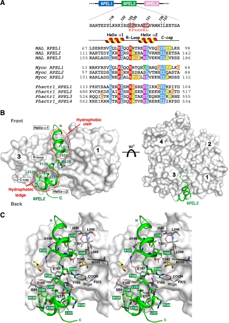
Serum response factor transcriptional activity is controlled through interactions with regulatory cofactors such as the coactivator MAL/MRTF-A (myocardin-related transcription factor A). MAL is itself regulated in vivo by changes in cellular actin dynamics, which alter its interaction with G-actin. The G-actin-sensing mechanism of MAL/MRTF-A resides in its N-terminal domain, which consists of three tandem RPEL repeats. We describe the first molecular insights into RPEL function obtained from structures of two independent RPEL(MAL) peptide:G-actin complexes. Both RPEL peptides bind to the G-actin hydrophobic cleft and to subdomain 3. These RPEL(MAL):G-actin structures explain the sequence conservation defining the RPEL motif, including the invariant arginine. Characterisation of the RPEL(MAL):G-actin interaction by fluorescence anisotropy and cell reporter-based assays validates the significance of actin-binding residues for proper MAL localisation and regulation in vivo. We identify important differences in G-actin engagement between the two RPEL(MAL) structures. Comparison with other actin-binding proteins reveals an unexpected similarity to the vitamin-D-binding protein, extending the G-actin-binding protein repertoire.


Figures

Figure 1
Figure 1
Structure of an RPEL peptide bound to G-actin. (A) Sequence alignment of individual RPEL motifs from murine MAL, myocardin (transcript variant A) and Phactr1. RPEL2MAL secondary structure and features discussed in text are shown above the sequence. Selected conserved residues are highlighted. (B) Two views of the RPEL2MAL:G-actin complex, related by a 90° rotation around the horizontal axis. Right-hand panel is the classical view of the ‘front' surface of actin (white with subdomains labelled 1–4). RPEL2MAL is drawn in green (cartoon) with highly conserved RPEL residues that interact with actin shown as sticks. The hydrophobic cleft and the subdomain 3 ledge of actin are indicated by red dashed circles. (C) Stereo view of the RPEL2MAL (green cartoon) interaction with G-actin. Actin surface is drawn as per (B) with selected RPEL-interacting residues shown as grey sticks and key hydrogen bonds are indicated as dashed lines. Two glycerol molecules, used as a cryoprotectant, are shown in yellow.
Figure 2
Figure 2
The RPEL1MAL and RPEL2MAL R-loops make different contacts with G-actin. (A) Summary of RPEL1/RPEL2 interactions with G-actin mapped onto a HMM (Hidden Markov Model) representation for the RPEL motif (Schuster-Bockler et al, 2004). Helices α1 and α2 are highlighted in pink and conserved residues from the RPEL HMM are highlighted in yellow. Boxes with solid lines indicate RPELMAL side chain-mediated interactions with actin, whereas those with dashed lines describe RPELMAL main chain-mediated interactions. Interactions that are conserved between RPEL peptides 1 and 2 are shown in black text and RPEL1- and RPEL2-specific ones in blue and green, respectively. (B) Close-up view of RPEL1MAL and RPEL2MAL interactions close to Y169actin. Left panel, RPEL1MAL:G-actin; right panel, RPEL2MAL:G-actin. A 2mFoDFc electron density map calculated around each RPEL is shown in blue contoured at 1 σ. Note that F375 is disordered in the RPEL1:actin complex. (C) Comparison of RPEL1MAL (cyan) and RPEL2MAL (green) motifs following superposition of their respective actin subunits. Important RPEL and G-actin residues described in the text are highlighted. Selected actin residues are shown in dark blue (contacting RPEL1) and dark green (contacting RPEL2), respectively. (D) Loss of F375 of β-actin affects binding of MAL RPEL motifs differentially. NIH3T3 fibroblasts transiently expressing either wild-type FLAG–β-actin (W) or FLAG–β-actin-ΔF375 (Δ) lacking the C-terminal residue were lysed and extracts were probed with bacterially produced GST or GST–RPEL peptide fusions as indicated. NIH3T3 cell lysates (input) and bound material were subjected to SDS–PAGE and western blotting for detection of the FLAG tag (WB: anti-FLAG) or endogenous β-actin (WB: anti-β-actin). Ponceau stain of the membrane indicates the levels of GST fusion proteins.
Figure 3
Figure 3
In vitro and in vivo validation of the RPEL1MAL and RPEL2MAL structures. (A) Fluorescence anisotropy assay for characterisation of the RPELMAL:G-actin interaction. Anisotropies of FITC-conjugated 32 amino-acid RPEL peptides at a concentration of 0.5 μM were measured over a range of LatB–actin concentrations. Anisotropy values were normalised by subtracting the anisotropy obtained in the absence of LatB–actin from all anisotropies for each peptide and multiplied by 1000. Graphs correspond to one of three experiments done in duplicate. Dissociation constants (Kd) for RPELMAL:G-actin interactions were calculated by nonlinear regression from each duplicate after normalisation using GraFit software (see Materials and methods). Kd values shown are means from three independent experiments with s.e.m. (B) Schematic representation of N-terminal MAL mutations used for luciferase reporter assays and immunofluorescence. The mutated region is shown in red. (C) SRF reporter activation by structure-derived MAL point mutants. The indicated MAL derivatives were expressed with and without C3 transferase coexpression in serum-starved NIH3T3 cells. Reporter activation was normalised to reporter activation conferred by SRF-VP16 or SRF-VP16 plus C3 transferase. x23, 1x3, 12x and xxx refer to MAL derivatives described earlier (Guettler et al, 2008): x23, R81A; 1x3, R125A; 12x, R169A; xxx, R81A R125A R169A. Data from three independent experiments are shown. Error bars, s.e.m. (D) Subcellular localisation of structure-derived MAL point mutants. The localisation of the indicated constructs was scored as predominantly nuclear (nuc), comparable intensity in nucleus and cytoplasm (nuc/cyt) or predominantly cytoplasmic (cyt) in 100 serum-starved cells. Mutants are described in (B,C). Data from three independent experiments are shown. Error bars, s.e.m.
Figure 4
Figure 4
Structural comparison with known G-actin-binding proteins. (A) Left: the DBP:G-actin complex structure (PDB code 1MA9, actin as a white surface, DBP as grey cartoon ribbon). DBP helices that interact with the actin hydrophobic cleft and subdomain 3 ledge are shown in orange. Right: close-up of the actin hydrophobic cleft showing the binding interface of DBP and RPEL2MAL superposed onto their respective G-actin partners. RPEL2MAL is shown in green. DBP residue numbering is taken from the 1MA9 coordinates. (B) Left: bottom view of superposed WH2 motif containing proteins together with RPEL2MAL bound to G-actin. MIM, black (PDB code 2D1K); WIP, marine blue (2A41); WASP, yellow (2A3Z); WAVE2, pink (2A40); Ciboulot, red (1SQK); RPEL2MAL (green) gelsolin, purple (1EQY). Right: close up of the left-hand panel showing the side chains at the four conserved positions (A–D) together with an electrostatic surface of actin (red indicating acidic regions). The RPEL2MAL α1 helix and its residue numbers are shown in green. (C) Structural alignment of helices equivalent to RPEL helix α1 from various G-actin-binding proteins. The four equivalent key residues common to these actin-binding proteins are displayed as sticks. The orientation of each helix is indicated in parentheses; R, reverse and F, forward.
Figure 5
Figure 5
Structure-based sequence alignment of G-actin-binding proteins engaging the subdomain 1–3 hydrophobic cleft. The alignment is subdivided according to forward and reverse orientations of the actin-binding α-helix. Red boxes indicate experimentally observed helical regions. Cleft-binding residues A, B, C and D are highlighted as well as the three actin ledge-binding residues. Residue numbering for gelsolin and DBP are taken from the PDB coodinate files indicated and can be interconverted to full length sequence numbering by addition of 51 or 16 residues respectively.

References

    1. Allen PB, Greenfield AT, Svenningsson P, Haspeslagh DC, Greengard P (2004) Phactrs 1–4: a family of protein phosphatase 1 and actin regulatory proteins. Proc Natl Acad Sci USA 101: 7187–7192 - PMC - PubMed
    1. Bubb MR, Govindasamy L, Yarmola EG, Vorobiev SM, Almo SC, Somasundaram T, Chapman MS, Agbandje-McKenna M, McKenna R (2002) Polylysine induces an antiparallel actin dimer that nucleates filament assembly: crystal structure at 3.5-Å resolution. J Biol Chem 277: 20999–21006 - PubMed
    1. Burtnick LD, Urosev D, Irobi E, Narayan K, Robinson RC (2004) Structure of the N-terminal half of gelsolin bound to actin: roles in severing, apoptosis and FAF. EMBO J 23: 2713–2722 - PMC - PubMed
    1. CCP4 (Collaborative Computational Project N) (1994) The CCP4 suite: programs for protein crystallography. Acta Crystallogr D Biol Crystallogr 50: 760–763 - PubMed
    1. Cen B, Selvaraj A, Burgess RC, Hitzler JK, Ma Z, Morris SW, Prywes R (2003) Megakaryoblastic leukemia 1, a potent transcriptional coactivator for serum response factor (SRF), is required for serum induction of SRF target genes. Mol Cell Biol 23: 6597–6608 - PMC - PubMed

Publication types

MeSH terms