

Review
doi: 10.1016/j.ymeth.2008.10.024.
Epub 2008 Nov 14.
Oligonucleotide-based assays for integrase activity
Affiliations
- PMID: 19010419
- PMCID: PMC2743288
- DOI: 10.1016/j.ymeth.2008.10.024
Item in Clipboard
Review
Oligonucleotide-based assays for integrase activity
Methods.
2009 Apr.
Abstract
Oligonucleotide assays have been invaluable for elucidation of the molecular mechanisms of retroviral integrases. A suite of rapid and sensitive fluorescence assays to measure the DNA binding, processing, and joining activities of integrase (IN) is described here. The assays are especially useful for characterizing the major activities of the enzyme, and for handling large numbers of samples efficiently. They can greatly facilitate further biochemical and structural analyses for HIV-1 and other IN proteins. The assays can also be adapted for moderate-high throughput testing of various inhibitory compounds.
Figures
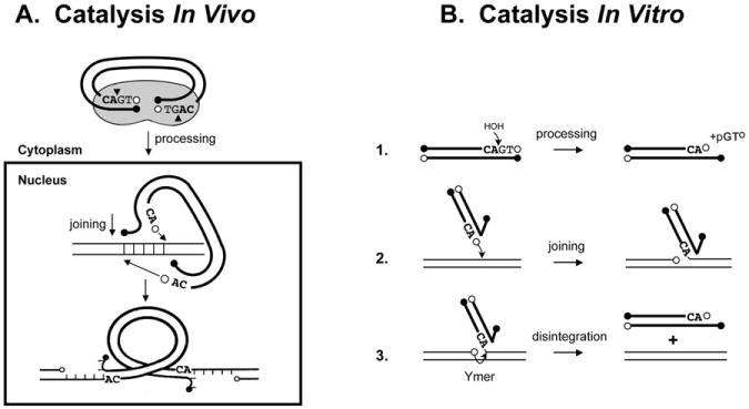
Catalysis by retroviral integrase (IN). (A), Shows the steps known to occur in vivo. (B), Shows the steps that can be measured separately in vitro using short oligodeoxyribonucleotide duplexes (oligos) that represent a viral DNA end (heavy lines) and a target DNA (light lines). The last four nucleotides in the 3′-ends of HIV-1 DNA are shown. Open circles indicate 3′-OH ends; filled circles, 5′-PO4 ends.
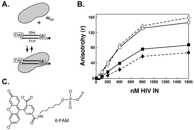
Binding of HIV-1 IN to a 6-FAM-labeled, recessed viral DNA oligo. (A), Illustration of the reaction. (B), Anisotropy measurements. Diamonds (dashed lines) show results with 6-His-tagged IN; solid lines are results with untagged IN. Solid symbols show binding in the presence of MgCl2; open symbols, in the presence of MnCl2. (C), Structure of 6-carbofluorescein (6-FAM). Conjugation occurs through the terminal phosphate.
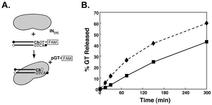
Processing by 6-His-tagged or untagged HIV-1 IN in the presence of MgCl2. (A), Illustration of the reaction. (B), Diamonds (dashed lines) show product released with 6-His-tagged IN, and squares (solid line) with untagged IN.
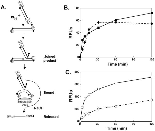
Fluorescent joining assay of 6-His-tagged or untagged HIV-1 IN in the presence of MgCl2 or MnCl2. (A), Illustrates the single end joining assay in which the product is bound to streptavidin-coated beads via the biotin-conjugated target oligo. The bound FAM-labeled viral strand is released upon denaturation by addition of NaOH. The amount of released, FAM-labeled viral strand is then quantified. (B), Joining activity as a function of time in the presence of MgCl2. (C), Joining activity as a function of time in the presence of MnCl2. Note the 10-fold increase in scale compared with (B). In this assay 400 RFU ≃1% joining of viral oligo. Diamonds (dashed lines) represent data points with 6-His-tagged IN; squares (solid lines) with untagged IN.
References
Publication types
MeSH terms
Substances
Grants and funding
LinkOut - more resources
Full Text Sources
Other Literature Sources
