. 2009 Feb;44(2):275-86.
doi: 10.1016/j.bone.2008.10.037. Epub 2008 Oct 22.

Intermittent PTH stimulates periosteal bone formation by actions on post-mitotic preosteoblasts

Affiliations

Intermittent PTH stimulates periosteal bone formation by actions on post-mitotic preosteoblasts

Robert L Jilka et al. Bone. 2009 Feb.

Abstract

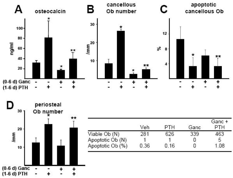
Intermittent administration of parathyroid hormone (PTH) stimulates bone formation on the surface of cancellous and periosteal bone by increasing the number of osteoblasts. Previous studies of ours in mice demonstrated that intermittent PTH increases cancellous osteoblast number at least in part by attenuating osteoblast apoptosis, but the mechanism responsible for the anabolic effect of the hormone on periosteal bone is unknown. We report that daily injections of 100 ng/g of PTH(1-34) to 4-6 month old mice increased the number of osteoblasts on the periosteum of lumbar vertebrae by 2-3 fold as early as after 2 days. However, the prevalence of apoptotic periosteal osteoblasts was only 0.2% in vehicle treated animals, which is approximately 20-fold lower than is the case for cancellous osteoblasts. Moreover, PTH did not have a discernable effect on periosteal osteoblast apoptosis. Administration of BrdU for 4 days failed to label periosteal osteoblasts under either basal conditions or following administration of PTH. Cancellous osteoblasts, on the other hand, were labeled under basal conditions, but PTH did not increase the percentage of BrdU-positive cells. Thus, intermittent PTH does not increase cancellous or periosteal osteoblast number by stimulating the proliferation of osteoblast progenitors. Consistent with high turnover of cancellous osteoblasts as compared to that of periosteal osteoblasts, ganciclovir-induced ablation of replicating osteoblast progenitors in mice expressing thymidine kinase under the control of the 3.6 kb rat Col1A1 promoter resulted in disappearance of osteoblasts from cancellous bone over a 7-14 day period, whereas periosteal osteoblasts were unaffected. However, 14 days of pre-treatment with ganciclovir prevented PTH anabolism on periosteal bone. We conclude that in cancellous bone, attenuation of osteoblast apoptosis by PTH increases osteoblast number because their rate of apoptosis is high, making this effect of the hormone profound. However, in periosteal bone where the rate of osteoblast apoptosis is low, PTH must exert pro-differentiating and/or pro-survival effects on post-mitotic pre-osteoblasts. Targeting the latter cells is an effective mechanism for increasing osteoblast number in periosteal bone where the production of osteoblasts from replicating progenitors is slow.


Figures

Figure 1
Figure 1. The stimulatory effect of intermittent PTH on periosteal bone formation is not associated with attenuation of osteoblast apoptosis
(A–C) Female Swiss-Webster mice were injected daily with 100 ng/g PTH, or vehicle (Veh), for 28 days (N=5 per group). Nondecalcified sections of lumbar vertebrae were used to determine (A) the number of periosteal osteoblasts (Ob) visualized by staining with toluidene blue (arrows), (B) tetracycline-based bone formation rate, and (C) the number of apoptotic periosteal osteoblasts visualized with ISEL-staining. Arrow head, ISEL-labeled periosteal osteoblast; black arrows, viable periosteal osteoblasts; red arrows viable endosteal and cancellous osteoblasts. (A–C) b, cortical bone; original magnification = 400X. (D) Female Swiss-Webster mice were injected daily with 100 ng/g PTH for the indicated number of days, or left untreated (N=5 per group), and periosteal osteoblast number in lumbar vertebrae determined. *P<0.05 vs. vehicle-treated (A,B) or untreated (D) animals. (C,D) There was no statistically significant effect of PTH on the prevalence of periosteal osteoblast apoptosis as determined by Poisson test of homogeneity.
Figure 2
Figure 2. The PTH-stimulated increase in osteoblast number on cancellous and periosteal bone is not due to increased osteoblast progenitor replication
(A) Left panel. BrdU labeling protocol used to obtained data shown in panels B and C. Groups of 5 month old female Swiss-Webster mice (indicated by bars, N=5–6 per group) were implanted with BrdU pellets at the indicated times during treatment with vehicle (Veh) or PTH (100 ng/g/d) by daily injection. Right panel. BrdU-labeled cancellous osteoblasts (arrows). b, cancellous bone; original magnification = 400X. (B) The total number of osteoblasts, and the number that were BrdU-labeled (BrdU+), on cancellous bone (left panel) or periosteal bone (right panel) were determined in decalcified sections of lumbar vertebrae. The number of BrdU-labeled periosteal osteoblasts in PTH-treated animals are too low to be depicted in the Figure (0.14 per mm the 48–96 h group, and the 0.03/mm in the 72–96 h group). (C) The percentage of cancellous osteoblasts labeled during each of the labeling intervals depicted in panel A was determined as follows. The percentage of BrdU-positive osteoblasts labeled during the 24–72 h period was subtracted from that labeled during the 0–96 h period to yield the percentage of osteoblasts that developed from progenitors that had divided during the first 24 h of the experiment, as indicated by the bracket in panel A. Similar calculations yield labeling indices for the 24–48 h and 48–72 h intervals. The 72–96 h labeling index was determined directly. * P<0.05 vs. respective vehicle control.
Figure 3
Figure 3. Characteristics of 3.6Col1A1-tk mice
(A) The 3.6Col-tk transgene construct contains 3.6 kb of the rat collagen 1A1 5′-flanking region upstream from the herpes virus thymidine kinase (HSV tk) cDNA. To provide a heterologous intron and polyadenylation site, a fragment containing an artificial intron (int) followed by an internal ribosome entry site-puromycin-resistance cassette (IRES-puro) and polyadenylation site (PA) was inserted downstream from the tk cDNA. (B) Expression of tk mRNA in bone and other tissues from 3.6Col1A1-tk mice, determined by qPCR. Data shown represent the mean of samples from 3 mice relative to GAPDH (ND = not detected). (C) Immunostaining for tk in osteoblasts (arrows), and osteocytes (arrow heads) in cancellous and periosteal bone of lumbar vertebrae. b, bone; bm, bone marrow, original magnification = 400X.
Figure 4
Figure 4. Effect of ganciclovir on 3.6Col1A1-tk mice
(A,B) 3.6Col1A1-tk mice (2 months old, N=5–7 per group) were given twice daily injections of vehicle (veh) or ganciclovir for 28 days. Serum osteocalcin level (A) was determined at the indicated times, and transcript levels of osteocalcin, Col1A1, Sost, and tk were determined in vertebral bone by qPCR (B) at the end of the experiment. (C) 3.6Col1A1-tk mice (4–5 months old, N=6 per group) were given twice daily injections of vehicle or ganciclovir for 14 days. Osteoblast (Ob) number and bone formation rate in cancellous bone of lumbar vertebrae were determined by histomorphometry. (D) 3.6Col1A1-tk mice (2 months old, N=5–7 per group) were given twice daily injections of vehicle or ganciclovir for 6 days. Osteoblasts (arrows) adjacent to osteoid in a nondecalcified section of vertebral cancellous bone after 6 days of ganciclovir administration. toluidene blue staining; original magnification 400X. b, bone; Apoptotic osteoblasts were quantified by ISEL staining in wild type and 3.6Col1A1-tk mice after 6 days of ganciclovir administration. (E) Femoral colony forming osteoblast progenitors were determined in ex-vivo cultures in 6-well plates after 14 days of vehicle or ganciclovir administration in the experiment shown in panel C. Mineralized matrix was visualized with Von Kossa staining. (F) Osteoblast number and indices of bone formation in periosteal bone of lumbar vertebrae determined by histomorphometry in the experiment shown in panel C. * P<0.05 vs. vehicle.
Figure 5
Figure 5. The PTH-stimulated increase in cancellous and periosteal osteoblasts is preserved when co-administered with ganciclovir for 6 days
Col1A1-tk mice (4 months old, N=6 per group) were injected twice daily with PBS or ganciclovir (Ganc) as in Figure 4 for 1 day, followed by 6 days of 100 ng/g PTH or vehicle injections while maintaining the ganciclovir administration protocol. (A) Circulating osteocalcin was determined at the end of the experiment. (B–D) Osteoblast (Ob) number and the prevalence of osteoblast apoptosis was detemined in cancellous and periosteal bone of lumbar vertebrae. * P<0.05 vs. mice receiving neither PTH nor ganciclovir; ** P<0.05 vs. mice receiving ganciclovir alone. There was no statistically significant difference in the prevalence of periosteal osteoblast apoptosis among the treatment groups as determined by Poisson test of homogeneity.
Figure 6
Figure 6. The increase in periosteal osteoblasts caused by 5 daily PTH injections is lost following pretreatment with ganciclovir for 14 days
Col1A1-tk mice (4 months old, N=5–7 per group) were injected twice daily for 14 days with PBS or ganciclovir (Ganc), followed by 5 days of 100 ng/g PTH, or vehicle, injections while maintaining the ganciclovir administration protocol. Periosteal osteoblast (Ob) number and the prevalence of periosteal osteoblast apoptosis was determined in lumbar vertebrae. * P<0.05 vs. mice receiving neither ganciclovir nor PTH. There was no statistically significant difference in the prevalence of periosteal osteoblast apoptosis among the treatment groups as determined by Poisson test of homogeneity.
Figure 7
Figure 7. The increase in periosteal osteoblasts and bone formation rate caused by 14 daily PTH injections is lost following pretreatment with ganciclovir for 14 days
Col1A1-tk mice (4 months old, N=5–7 per group) were injected twice daily for 14 days with PBS or ganciclovir (Ganc), followed by 14 days of 100 ng/g PTH, or vehicle, injections while maintaining the ganciclovir administration protocol. Periosteal osteoblast (Ob) number and the prevalence of periosteal osteoblast apoptosis (A), as well as indices of bone formation (B–E) were determined in lumbar vertebrae. * P<0.05 vs. mice receiving neither ganciclovir nor PTH. There was no statistically significant difference in the prevalence of periosteal osteoblast apoptosis among the treatment groups as determined by Poisson test of homogeneity.
Figure 8
Figure 8. Intermittent PTH stimulates bone formation in periosteal and cancellous bone by different mechanisms
Mitotic osteoblast progenitors give rise to post-mitotic preosteoblasts and eventually mature osteoblasts, which then die by apoptosis or become osteocytes or lining cells. Intermittent PTH exerts anti-apoptotic and/or pro-differentiating effects on post-mitotic preosteoblasts in periosteal bone, and anti-apoptotic effects on mature osteoblasts in cancellous bone. The result in both sites is an increase in the number of osteoblasts and thereby an increase in bone formation rate. See text for further details.

References

    1. Compston JE. Skeletal actions of intermittent parathyroid hormone: effects on bone remodelling and structure. Bone. 2007;40:1447–52. - PubMed
    1. Hodsman AB, Bauer DC, Dempster D, Dian L, Hanley DA, Harris ST, Kendler D, McClung MR, Miller PD, Olszynski WP, Orwoll E, Yuen CK. Parathyroid hormone and teriparatide for the treatment of osteoporosis: a review of the evidence and suggested guidelines for its use. Endocr Rev. 2005;26:688–703. - PubMed
    1. Neer RM, Arnaud CD, Zanchetta JR, Prince R, Gaich GA, Reginster JY, Hodsman AB, Eriksen EF, Ish-Shalom S, Genant HK, Wang O, Mitlak BH. Effect of parathyroid hormone (1–34) on fractures and bone mineral density in postmenopausal women with osteoporosis. N Engl J Med. 2001;344:1434–41. - PubMed
    1. Iida-Klein A, Lu SS, Cosman F, Lindsay R, Dempster DW. Effects of cyclic vs. daily treatment with human parathyroid hormone (1–34) on murine bone structure and cellular activity. Bone. 2007;40:391–8. - PubMed
    1. Jilka RL, Weinstein RS, Bellido T, Roberson P, Parfitt AM, Manolagas SC. Increased bone formation by prevention of osteoblast apoptosis with parathyroid hormone. J Clin Invest. 1999;104:439–46. - PMC - PubMed

Publication types

MeSH terms