Review
. 2009 Mar;5(3):817-31.
doi: 10.1016/j.actbio.2008.09.018. Epub 2008 Oct 10.

Self-assembling materials for therapeutic delivery

Affiliations
Review

Self-assembling materials for therapeutic delivery

Monica C Branco et al. Acta Biomater. 2009 Mar.

Abstract

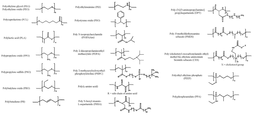
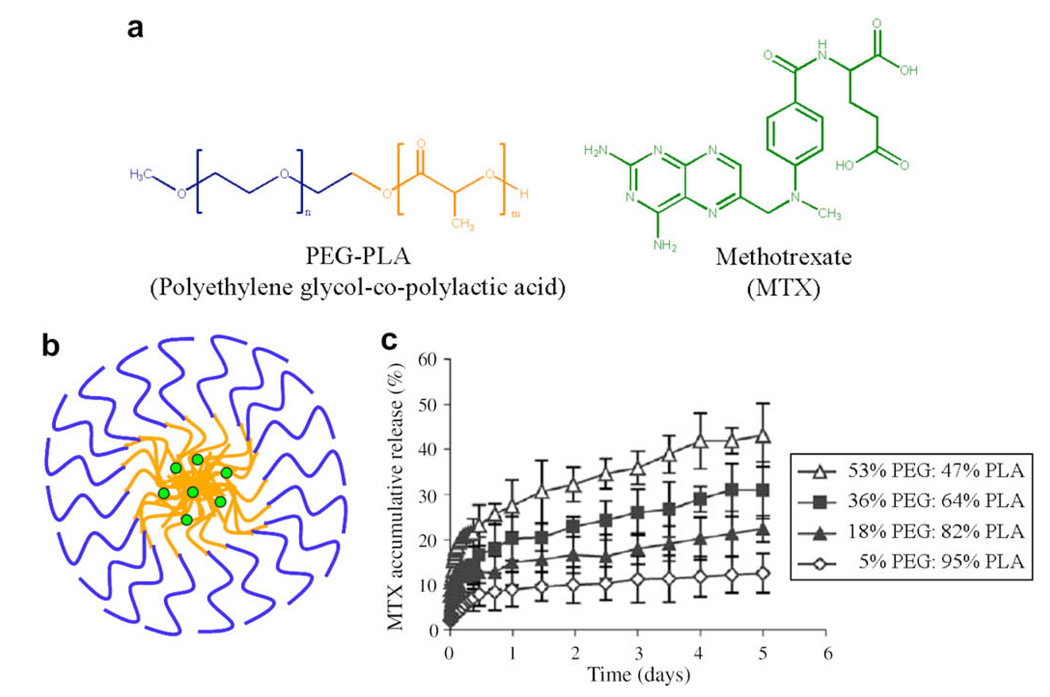
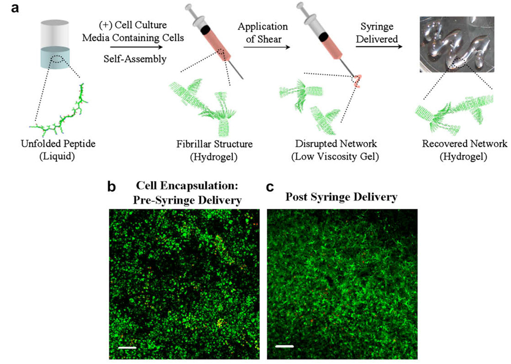
A growing number of medications must be administered through parenteral delivery, i.e., intravenous, intramuscular, or subcutaneous injection, to ensure effectiveness of the therapeutic. For some therapeutics, the use of delivery vehicles in conjunction with this delivery mechanism can improve drug efficacy and patient compliance. Macromolecular self-assembly has been exploited recently to engineer materials for the encapsulation and controlled delivery of therapeutics. Self-assembled materials offer the advantages of conventional crosslinked materials normally used for release, but also provide the ability to tailor specific bulk material properties, such as release profiles, at the molecular level via monomer design. As a result, the design of materials from the "bottom up" approach has generated a variety of supramolecular devices for biomedical applications. This review provides an overview of self-assembling molecules, their resultant structures, and their use in therapeutic delivery. It highlights the current progress in the design of polymer- and peptide-based self-assembled materials.


Figures

Fig 1
Fig 1
Common self-assembling monomers include lipids, block copolymers, peptides and proteins. Intermolecular interactions that drive and define self-assembly include hydrophobic association and the formation of polar interactions, respectively. The resultant structures formed through self-assembly are shown. The hydrophilic portions (blue) and hydrophobic portions (orange) have been color-coded. (For interpretation of color mentioned in this figure the reader is referred to the web version of the article.)
Fig 2
Fig 2
(a) Common therapeutics with sizes ranging from the nanoscale to the microscale. (b) Self-assembled structures used for therapeutic delivery, highlighting the types of encapsulated therapeutics and methods of release. In general, release is diffusion-controlled but systems can be engineered to undergo active degradation or disassembly. The hydrophilic portions (blue) and hydrophobic portions (orange) have been color-coded. (For interpretation of color mentioned in this figure the reader is referred to the web version of the article.)
Fig 3
Fig 3
Common polymers used for self-assembled delivery vehicles.
Fig 4
Fig 4
(a) Structure of polyethylene glycol–co-polylactic acid (PEG–PLA) and the anticancer drug methotrexate (MTX). (b) A sketch depicting the drug-loaded micelle. The hydrophilic portions (blue) and hydrophobic portions (orange) of the copolymer have been color-coded. (c) Release profiles of MTX from PEG–PLA micelles with varying weight per cents of block copolymers in phosphate-buffered saline (PBS) at 37°C. As the weight per cent of the hydrophobic polymer increases, the rate of delivery decreases. Reprinted from Ying Zhang et al., Methotrexate-loaded biodegradable polymeric micelles: preparation, physicochemical properties and in vitro drug release, Colloids and Surfaces B: Biointerfaces, 2004;44(2–3):104–109, with permission from Elsevier. (For interpretation of color mentioned in this figure the reader is referred to the web version of the article.)
Fig 5
Fig 5
(a) Structure of the block copolymers, polyethylene glycol–co-polybutadiene (PEG–PB) and PEG–polylactic acid (PEG–PLA). The hydrophilic and hydrophobic portions of PEG–PB are dark blue and dark orange, respectively; the hydrophilic and hydrophobic portions of PEG–PLA are light blue and light orange, respectively. The structures of the anticancer drugs, doxorubicin and paclitaxol, are shown. (b) Sketch of vesicle containing 75% PEG–PB and 25% PEG–PLA encapsulating doxorubicin in the aqueous lumen and paclitaxol in the hydrophobic bilayer. (c) Plot showing the relative tumor area as a function of time after a single injection of the indicated therapeutic. Tumors were formed by subcutaneous injection of MDA-MB231 cells (2 × 10⁶ cells initially) into nude mice. Therapeutic was delivered by tail-vein injection. Tumor areas are normalized to the tumor areas of the control groups: empty polymersomes and saline injection. No differences were seen between these two control groups. Reprinted from Fariyal Ahmed et al., Biodegradable polymersomes loaded with both paclitaxel and doxorubicin permeate and shrink tumors, inducing apoptosis, Journal of Controlled Release, 2006; 116(2):150–158, with permission from Elsevier. (For interpretation of color mentioned in this figure the reader is referred to the web version of the article.)
Fig 6
Fig 6
(a) Mechanism of self-assembly and shear-thinning properties of MAX8 peptide hydrogel. Under low ionic strength, aqueous conditions, the peptide is unfolded and freely soluble. Folding and assembly is triggered by the addition of cell culture media to yield a mechanically rigid, noncovalently crosslinked hydrogel directly within a syringe. Depressing the syringe plunger applies stress to the gel, converting it into a low-viscosity gel that flows and can be delivered through the syringe needle. At the site of delivery, the gel immediately reforms, recovering its mechanical rigidity. When gelation is triggered in the presence of cells, the cells become encapsulated and the resulting gel–cell construct can be delivered via syringe. Cells are not shown in (a) for clarity. (b) Live/dead cell viability assay showing C3H10t1/2 mesenchymal stem cells in 0.5 wt.% MAX8 hydrogels after triggered encapsulation in the gel. Image was taken two weeks after encapsulation. Live cells are green; dead cells are red. (c) Live/dead viability assay showing encapsulated mesenchymal stem cells after being shear-thin delivered with a syringe. Image taken two weeks after delivery. 50 × 106 cells were initially loaded into the scaffold. Scale bar is 100 µm. (For interpretation of color mentioned in this figure the reader is referred to the web version of the article.)

References

    1. Reichert JM. Trends in US approvals: new biopharmaceuticals and vaccines. Trends Biotechnol. 2006;24:293–298. - PMC - PubMed
    1. Reichert JM, Wenger JB. Development trends for new cancer therapeutics and vaccines. Drug Discov Today. 2008;13:30–37. - PubMed
    1. Chaubal MV, Roseman TJ. Drug delivery trends for parenteral therapeutics. Drug Deliv Syst. 2006;21:388–397.
    1. Pawar R, Ben-Ari A, Domb AJ. Protein and peptide parenteral controlled delivery. Exp Opin Biol Ther. 2004;4:1203–1212. - PubMed
    1. Sagar GH, Arunagirinathan MA, Bellare JR. Self-assembled surfactant nano-structures important in drug delivery: a review. Indian J Exp Biol. 2007;45:133–159. - PubMed

Publication types