

Phospholipid scramblases and Tubby-like proteins belong to a new superfamily of membrane tethered transcription factors
- PMID: 19010806
- PMCID: PMC2639001
- DOI: 10.1093/bioinformatics/btn595
Phospholipid scramblases and Tubby-like proteins belong to a new superfamily of membrane tethered transcription factors
Abstract
Motivation: Phospholipid scramblases (PLSCRs) constitute a family of cytoplasmic membrane-associated proteins that were identified based upon their capacity to mediate a Ca(2+)-dependent bidirectional movement of phospholipids across membrane bilayers, thereby collapsing the normally asymmetric distribution of such lipids in cell membranes. The exact function and mechanism(s) of these proteins nevertheless remains obscure: data from several laboratories now suggest that in addition to their putative role in mediating transbilayer flip/flop of membrane lipids, the PLSCRs may also function to regulate diverse processes including signaling, apoptosis, cell proliferation and transcription. A major impediment to deducing the molecular details underlying the seemingly disparate biology of these proteins is the current absence of any representative molecular structures to provide guidance to the experimental investigation of their function.
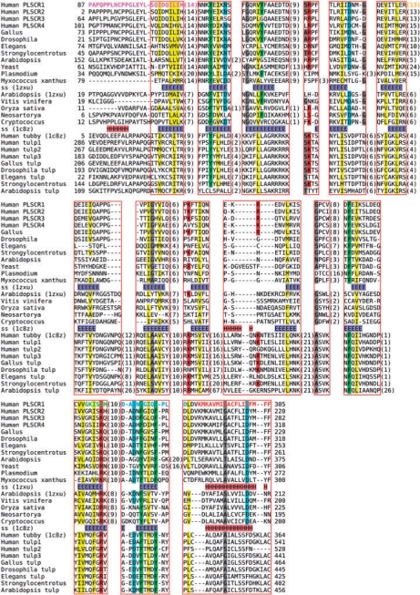
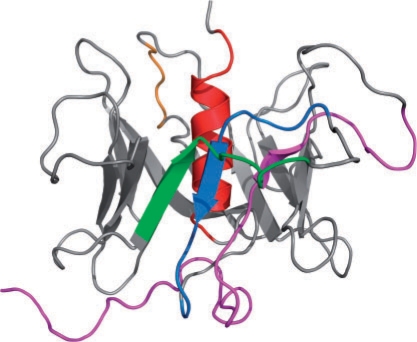
Results: Here, we show that the enigmatic PLSCR family of proteins is directly related to another family of cellular proteins with a known structure. The Arabidopsis protein At5g01750 from the DUF567 family was solved by X-ray crystallography and provides the first structural model for this family. This model identifies that the presumed C-terminal transmembrane helix is buried within the core of the PLSCR structure, suggesting that palmitoylation may represent the principal membrane anchorage for these proteins. The fold of the PLSCR family is also shared by Tubby-like proteins. A search of the PDB with the HHpred server suggests a common evolutionary ancestry. Common functional features also suggest that tubby and PLSCR share a functional origin as membrane tethered transcription factors with capacity to modulate phosphoinositide-based signaling.
Figures
References
-
- Amir-Moazami O, et al. Phospholipid scramblase 1 modulates a selected set of IgE receptor-mediated mast cell responses through LAT-dependent pathway. J. Biol. Chem. 2008;283:25514–25523. - PubMed
-
- Ben-Efraim I, et al. Phospholipid scramblase 1 is imported into the nucleus by a receptor-mediated pathway and interacts with DNA. Biochemistry. 2004;43:3518–3526. - PubMed
Publication types
MeSH terms
Substances
Grants and funding
LinkOut - more resources
Full Text Sources
Molecular Biology Databases
Miscellaneous
