Mutation of ERBB2 provides a novel alternative mechanism for the ubiquitous activation of RAS-MAPK in ovarian serous low malignant potential tumors
- PMID: 19010816
- PMCID: PMC6953412
- DOI: 10.1158/1541-7786.MCR-08-0193
Mutation of ERBB2 provides a novel alternative mechanism for the ubiquitous activation of RAS-MAPK in ovarian serous low malignant potential tumors
Abstract
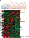
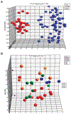
Approximately, 10% to 15% of serous ovarian tumors fall into the category designated as tumors of low malignant potential (LMP). Like their invasive counterparts, LMP tumors may be associated with extraovarian disease, for example, in the peritoneal cavity and regional lymph nodes. However, unlike typical invasive carcinomas, patients generally have a favorable prognosis. The mutational profile also differs markedly from that seen in most serous carcinomas. Typically, LMP tumors are associated with KRAS and BRAF mutations. Interrogation of expression profiles in serous LMP tumors suggested overall redundancy of RAS-MAPK pathway mutations and a distinct mechanism of oncogenesis compared with high-grade ovarian carcinomas. Our findings indicate that activating mutation of the RAS-MAPK pathway in serous LMP may be present in >70% of cases compared with approximately 12.5% in serous ovarian carcinomas. In addition to mutations of KRAS (18%) and BRAF (48%) mutations, ERBB2 mutations (6%), but not EGFR, are prevalent among serous LMP tumors. Based on the expression profile signature observed throughout our serous LMP cohort, we propose that RAS-MAPK pathway activation is a requirement of serous LMP tumor development and that other activators of this pathway are yet to be defined. Importantly, as few nonsurgical options exist for treatment of recurrent LMP tumors, therapeutic targeting of this pathway may prove beneficial, especially in younger patients where maintaining fertility is important.
Conflict of interest statement
Disclosure of Potential Conflicts of Interest
No potential conflicts of interest were disclosed.
Figures


References
-
- Stewart BW, Kleihues P, editors. IARC: World Cancer Report. Lyon: IARC Press; 2003.
-
- Choi KC, Auersperg N. The ovarian surface epithelium: simple source of a complex disease. Minerva Ginecol 2003;55:297–314. - PubMed
-
- Fukumoto M, Nakayama K. Ovarian epithelial tumors of low malignant potential: are they precursors of ovarian carcinoma? Pathol Int 2006;56:233–9. - PubMed
-
- Trimble EL, Trimble CL. Ovarian tumors of low malignant potential. Curr Treat Options Oncol 2001;2:103–8. - PubMed
-
- Sherman ME, Berman J, Birrer MJ, et al. Current challenges and opportunities for research on borderline ovarian tumors. Hum Pathol 2004;35:961–70. - PubMed
Publication types
MeSH terms
Substances
Grants and funding
LinkOut - more resources
Full Text Sources
Other Literature Sources
Medical
Molecular Biology Databases
Research Materials
Miscellaneous

