. 2008 Nov;6(11):1755-65.
doi: 10.1158/1541-7786.MCR-08-0095.

Mesothelin-induced pancreatic cancer cell proliferation involves alteration of cyclin E via activation of signal transducer and activator of transcription protein 3

Affiliations

Mesothelin-induced pancreatic cancer cell proliferation involves alteration of cyclin E via activation of signal transducer and activator of transcription protein 3

Uddalak Bharadwaj et al. Mol Cancer Res. 2008 Nov.

Abstract

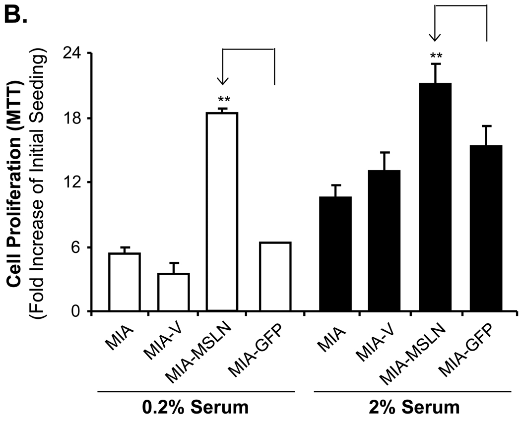
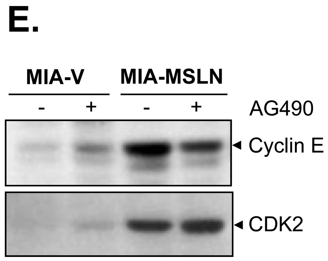
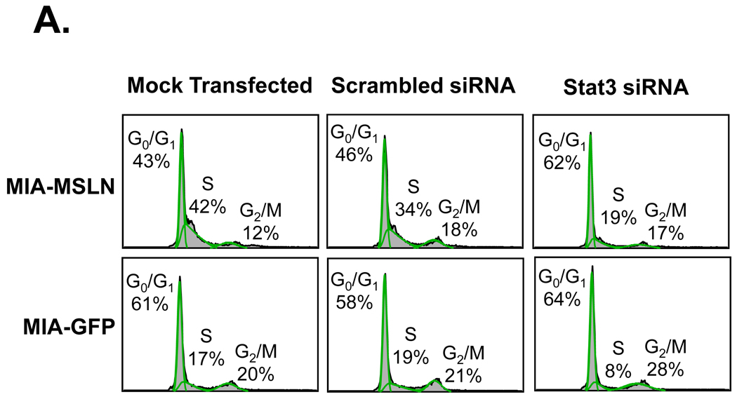
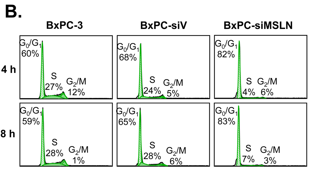
Mesothelin (MSLN) is a cell surface glycoprotein that is overexpressed in human pancreatic cancer. Although its value as a tumor marker for diagnosis and prognosis and as a preferred target of immunointervention has been evaluated, there is little information on the growth advantage of MSLN on tumor cells. In this study, we examined the effect of MSLN on pancreatic cancer cell proliferation, cell cycle progression, expression of cell cycle regulatory proteins, and signal transduction pathways in two pancreatic cancer cell lines, MIA-MSLN (overexpressing MSLN in MIA PaCa-2 cells) and BxPC-siMSLN (silencing MSLN in BxPC-3 cells). Increased cyclin E and cyclin-dependent kinase 2 expression found in MIA-MSLN cells correlated with significantly increased cell proliferation and faster cell cycle progression compared with control cells. BxPC-siMSLN cells showed slower proliferation and slower entry into the S phase than control cells. Signal transducer and activator of transcription protein 3 (Stat3) was constitutively activated in MIA-MSLN cells, but not in control cells. Inhibition of Stat3 activation in MIA-MSLN cells by the Janus-activated kinase-selective inhibitor tyrphostin AG490 was followed by a marked decrease in proliferation of the cells. Small interfering RNA against Stat3 significantly reduced the MIA-MSLN cell cycle progression with a concomitant decrease in cyclin E expression. Our data indicate that overexpression of MSLN in pancreatic cancer cells leads to constitutive activation of the transcription factor Stat3, which results in enhanced expression of cyclin E and cyclin E/cyclin-dependent kinase 2 complex formation as well as increased G(1)-S transition.


Conflict of interest statement

Disclosure of Potential Conflicts of Interest

The authors do not have potential conflicts of interest.

Figures

Figure 1
Figure 1
Overexpression of MSLN promotes pancreatic cancer cell proliferation and cell cycle progression. A. Cell proliferation of MIA-PaCa-2 cells according to MTT assay. MIA-MSLN and control cells were seeded in 96-well plates (2×10³ cells/well), serum-starved (0% FBS) for 24 h before changing to 2% FBS growth medium, and cultured for 6 d. Viability was measured with MTT. Relative increase in viability was measured by dividing viability at some time by viability of same cell at day 0 (day of addition of growth medium after initial serum starvation) and is plotted along Y axis. Data plotted show mean of triplicate wells. B. Serum dependence of proliferation. After initial 24 h of serum starvation, cells were treated with 0.2% and 2% serum growth medium; viability was measured with MTT after 3 d. C. Cell proliferation in Panc-1 cells (MTT assay). Panc1-MSLN and control cells were serum-starved (0% FBS) for 24 h before changing to 2% FBS growth medium, and cultured for 6 d. D. Cell cycle analysis. After initial 24 h of serum starvation and then release by 2% serum growth medium for 4 and 8 h, cells were collected, fixed, PI-stained, and analyzed for cell cycle phase distribution (percentage of cells) with FACS. E. Plating efficiency. MIA-V and MIA-MSLN cells (600) were plated in 150-mm dishes, allowed adhering for 48 h, and starved for 24 h. Cells were then allowed to form colonies in complete medium for 15 d, which were then stained with MTT. Percentage plating efficiency was determined as (number of colonies formed/cells seeded) × 100. Columns, mean of replicates; bars, SD; ** p < 0.001 compared with controls according to t test.
Figure 1
Figure 1
Overexpression of MSLN promotes pancreatic cancer cell proliferation and cell cycle progression. A. Cell proliferation of MIA-PaCa-2 cells according to MTT assay. MIA-MSLN and control cells were seeded in 96-well plates (2×10³ cells/well), serum-starved (0% FBS) for 24 h before changing to 2% FBS growth medium, and cultured for 6 d. Viability was measured with MTT. Relative increase in viability was measured by dividing viability at some time by viability of same cell at day 0 (day of addition of growth medium after initial serum starvation) and is plotted along Y axis. Data plotted show mean of triplicate wells. B. Serum dependence of proliferation. After initial 24 h of serum starvation, cells were treated with 0.2% and 2% serum growth medium; viability was measured with MTT after 3 d. C. Cell proliferation in Panc-1 cells (MTT assay). Panc1-MSLN and control cells were serum-starved (0% FBS) for 24 h before changing to 2% FBS growth medium, and cultured for 6 d. D. Cell cycle analysis. After initial 24 h of serum starvation and then release by 2% serum growth medium for 4 and 8 h, cells were collected, fixed, PI-stained, and analyzed for cell cycle phase distribution (percentage of cells) with FACS. E. Plating efficiency. MIA-V and MIA-MSLN cells (600) were plated in 150-mm dishes, allowed adhering for 48 h, and starved for 24 h. Cells were then allowed to form colonies in complete medium for 15 d, which were then stained with MTT. Percentage plating efficiency was determined as (number of colonies formed/cells seeded) × 100. Columns, mean of replicates; bars, SD; ** p < 0.001 compared with controls according to t test.
Figure 2
Figure 2
S-phase cyclin E and its binding partner CDK2 are up-regulated in MIA-MSLN cells. A. Sub-confluent cells were used to prepare lysates, and 60 µg of protein were subjected to SDS-PAGE and Western blot. Various cell-cycle-related proteins were detected with antibodies mentioned in methods.B. Control cells and MIA-MSLN cells were serum-starved (0% FBS) for 24 h, changed to 2% FBS medium, and collected at indicated times, and whole cell proteins were subjected to SDS PAGE, Western blotting, and probing for cyclin E, CDK2, and β actin. C. 400 µg of MIA-V and MIA-MSLN proteins was used to immunoprecipitate CDK2 by using immobilized protein G-conjugated-anti-CDK2 monoclonal antibody, and the immune-complex precipitate was washed and subjected to SDS-PAGE under denaturing conditions, gel-transferred to nitrocellulose membrane, and probed for cyclin E and CDK2.
Figure 2
Figure 2
S-phase cyclin E and its binding partner CDK2 are up-regulated in MIA-MSLN cells. A. Sub-confluent cells were used to prepare lysates, and 60 µg of protein were subjected to SDS-PAGE and Western blot. Various cell-cycle-related proteins were detected with antibodies mentioned in methods.B. Control cells and MIA-MSLN cells were serum-starved (0% FBS) for 24 h, changed to 2% FBS medium, and collected at indicated times, and whole cell proteins were subjected to SDS PAGE, Western blotting, and probing for cyclin E, CDK2, and β actin. C. 400 µg of MIA-V and MIA-MSLN proteins was used to immunoprecipitate CDK2 by using immobilized protein G-conjugated-anti-CDK2 monoclonal antibody, and the immune-complex precipitate was washed and subjected to SDS-PAGE under denaturing conditions, gel-transferred to nitrocellulose membrane, and probed for cyclin E and CDK2.
Figure 3
Figure 3
Blocking of Stat3 activation in MSLN-overexpressed MIA PaCa-2 cells led to decrease in proliferation and cell cycle progression. A. Activation of Stat3 in MIA-MSLN cells. 60 µg of total proteins from control and MIA-MSLN cells was subjected to immunoblot analysis with antibodies against phosphorylated form of Stat3 (pStat3Tyr705) and total Stat3. B. Nuclear translocation of Stat3 in MIA-MSLN cells. Nuclear protein was isolated from MIA-MSLN and control cells and subjected to SDS-PAGE and Western blot and probed for total Stat3 protein and nuclear envelope marker Lamin A as loading control. C. Blocking of Stat3 physophorylation with JAK-selective inhibitor Tyrphostin AG490. Total lysates from cells treated with Tyrphostin AG490 for 0, 4, 8, or 12 h were used to immunoblot for relative amount of pStat3Tyr705 and Stat3. D. Cell proliferation according to MTT assay. For cell-proliferation assay, control and MIA-MSLN cells were serum-starved for 24 h, treated with DMSO/Tyrphostin AG490 (50 µM) for 24 h in 2% serum medium, and washed. Proliferation was continued for 6 d, and cell viability was assayed with MTT. Cell proliferation of MIA-MSLN cells was significantly reduced by pretreatment with Tyrphostin AG490 (*p<0.05, ***p < 0.001). E. Effect of AG490 treatment on expression of cyclin E and CDK2. Whole proteins from MIA-V and MIA-MSLN cells, untreated or treated with AG490 for 48 h, were used to detect cyclin E and CDK2 with Western blot.
Figure 3
Figure 3
Blocking of Stat3 activation in MSLN-overexpressed MIA PaCa-2 cells led to decrease in proliferation and cell cycle progression. A. Activation of Stat3 in MIA-MSLN cells. 60 µg of total proteins from control and MIA-MSLN cells was subjected to immunoblot analysis with antibodies against phosphorylated form of Stat3 (pStat3Tyr705) and total Stat3. B. Nuclear translocation of Stat3 in MIA-MSLN cells. Nuclear protein was isolated from MIA-MSLN and control cells and subjected to SDS-PAGE and Western blot and probed for total Stat3 protein and nuclear envelope marker Lamin A as loading control. C. Blocking of Stat3 physophorylation with JAK-selective inhibitor Tyrphostin AG490. Total lysates from cells treated with Tyrphostin AG490 for 0, 4, 8, or 12 h were used to immunoblot for relative amount of pStat3Tyr705 and Stat3. D. Cell proliferation according to MTT assay. For cell-proliferation assay, control and MIA-MSLN cells were serum-starved for 24 h, treated with DMSO/Tyrphostin AG490 (50 µM) for 24 h in 2% serum medium, and washed. Proliferation was continued for 6 d, and cell viability was assayed with MTT. Cell proliferation of MIA-MSLN cells was significantly reduced by pretreatment with Tyrphostin AG490 (*p<0.05, ***p < 0.001). E. Effect of AG490 treatment on expression of cyclin E and CDK2. Whole proteins from MIA-V and MIA-MSLN cells, untreated or treated with AG490 for 48 h, were used to detect cyclin E and CDK2 with Western blot.
Figure 4
Figure 4
Stat3 siRNA treatment decreases normally increased cell cycle progression of MIA-MSLN cells. A. MIA-MSLN or MIA-GFP cells were transfected with either nonspecific scrambled siRNA oligonucleotide or Stat3-specific RNA pool. Cells treated with only transfection reagent was mock transfection control. For cell cycle analysis, 24 h after transfection, cells were serum-starved for 24 h, released with 2% serum medium, collected after 8 h, and processed for cell cycle analysis. B. Stat3 silencing decreased cyclin E expression in MIA-MSLN cells. Whole proteins from cells collected 48 h after transfection with Stat3-specific siRNA pool or scrambled siRNA control or transfection reagent control were used for Western blot with Stat3, cyclin E, and β actin antibodies.
Figure 4
Figure 4
Stat3 siRNA treatment decreases normally increased cell cycle progression of MIA-MSLN cells. A. MIA-MSLN or MIA-GFP cells were transfected with either nonspecific scrambled siRNA oligonucleotide or Stat3-specific RNA pool. Cells treated with only transfection reagent was mock transfection control. For cell cycle analysis, 24 h after transfection, cells were serum-starved for 24 h, released with 2% serum medium, collected after 8 h, and processed for cell cycle analysis. B. Stat3 silencing decreased cyclin E expression in MIA-MSLN cells. Whole proteins from cells collected 48 h after transfection with Stat3-specific siRNA pool or scrambled siRNA control or transfection reagent control were used for Western blot with Stat3, cyclin E, and β actin antibodies.
Figure 5
Figure 5
Silencing MSLN expression decreases pancreatic cancer cell proliferation and cell cycle progression. A. Cell proliferation according to MTT assay. MSLN siRNA-silenced BxPC-3 stable cell line (BxPC-siMSLN) and control cells BxPC-3 parental cells (BxPC-3) and empty siRNA-vector-integrated stable cell line (BxPC-siV) were seeded in 96-well plates (2×10³ cells/well) and serum-starved (0% FBS) for 24 h before being changed to growth medium with 2% FBS and cultured for 6 d. Cell growth was assessed at 2, 4, and 6 d after growth-medium addition with MTT assay. Cell proliferation of BxPC-siMSLN cells was significantly reduced compared with parental BxPC-3 and BxPC-siV cells (***p < 0.001). B. After initial serum starvation for 24 h, BxPC-siMSLN cells and controls were treated with 2% serum medium; cells were collected after 4 and 8 h, fixed, PI-stained, and analyzed for cell cycle phase distribution (percentage of cells) with FACS. C. Cyclin A and CDK2 expression was decreased in BxPC-siMSLN cells. Whole proteins from sub-confluent BxPC-siMSLN and control cells were subjected to SDS-PAGE and immunoblot for cell-cycle-related proteins.
Figure 5
Figure 5
Silencing MSLN expression decreases pancreatic cancer cell proliferation and cell cycle progression. A. Cell proliferation according to MTT assay. MSLN siRNA-silenced BxPC-3 stable cell line (BxPC-siMSLN) and control cells BxPC-3 parental cells (BxPC-3) and empty siRNA-vector-integrated stable cell line (BxPC-siV) were seeded in 96-well plates (2×10³ cells/well) and serum-starved (0% FBS) for 24 h before being changed to growth medium with 2% FBS and cultured for 6 d. Cell growth was assessed at 2, 4, and 6 d after growth-medium addition with MTT assay. Cell proliferation of BxPC-siMSLN cells was significantly reduced compared with parental BxPC-3 and BxPC-siV cells (***p < 0.001). B. After initial serum starvation for 24 h, BxPC-siMSLN cells and controls were treated with 2% serum medium; cells were collected after 4 and 8 h, fixed, PI-stained, and analyzed for cell cycle phase distribution (percentage of cells) with FACS. C. Cyclin A and CDK2 expression was decreased in BxPC-siMSLN cells. Whole proteins from sub-confluent BxPC-siMSLN and control cells were subjected to SDS-PAGE and immunoblot for cell-cycle-related proteins.

References

    1. Hassan R, Bera T, Pastan I. Mesothelin: a new target for immunotherapy. Clin Cancer Res. 2004;10:3937–3942. - PubMed
    1. Chang K, Pai LH, Batra JK, Pastan I, Willingham MC. Characterization of the antigen (CAK1) recognized by monoclonal antibody K1 present on ovarian cancers and normal mesothelium. Cancer Res. 1992;52:181–186. - PubMed
    1. Argani P, Iacobuzio-Donahue C, Ryu B, et al. Mesothelin is overexpressed in the vast majority of ductal adenocarcinomas of the pancreas: identification of a new pancreatic cancer marker by serial analysis of gene expression (SAGE) Clin Cancer Res. 2001;7:3862–3868. - PubMed
    1. Ho M, Bera TK, Willingham MC, et al. Mesothelin expression in human lung cancer. Clin Cancer Res. 2007;13:1571–1575. - PubMed
    1. Chang K, Pastan I. Molecular cloning of mesothelin, a differentiation antigen present on mesothelium, mesotheliomas, and ovarian cancers. Proc Natl Acad Sci U S A. 1996;93:136–140. - PMC - PubMed

Publication types

MeSH terms