. 2008 Nov 25;105(47):18171-5.
doi: 10.1073/pnas.0807296105. Epub 2008 Nov 14.

Nanoparticle-induced surface reconstruction of phospholipid membranes

Affiliations

Nanoparticle-induced surface reconstruction of phospholipid membranes

Bo Wang et al. Proc Natl Acad Sci U S A. .

Abstract

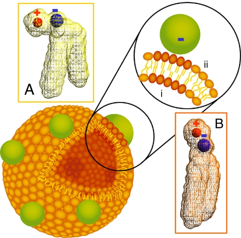
The nonspecific adsorption of charged nanoparticles onto single-component phospholipid bilayers bearing phosphocholine headgroups is shown, from fluorescence and calorimetry experiments, to cause surface reconstruction at the points where nanoparticles adsorb. Nanoparticles of negative charge induce local gelation in otherwise fluid bilayers; nanoparticles of positive charge induce otherwise gelled membranes to fluidize locally. Through this mechanism, the phase state deviates from the nominal phase transition temperature by tens of degrees. This work generalizes the notions of environmentally induced surface reconstruction, prominent in metals and semiconductors. Bearing in mind that chemical composition in these single-component lipid bilayers is the same everywhere, this offers a mechanism to generate patchy functional properties in phospholipid membranes.


Conflict of interest statement

The authors declare no conflict of interest.

Figures

Fig. 1.
Fig. 1.
Schematic diagram of a phospholipid bilayer vesicle with bound nanoparticles. Binding of anionic nanoparticles to a lipid bilayer in the fluid phase causes the nanoparticle to template a gel phase in the place where the nanoparticle binds. Binding-induced reorientation of the phosphocholine (PC) head group causes lipids in the fluid phase to have lower density (A) than in the gel phase (B). In the PC head group, P and N+ are denoted by blue and red, respectively.
Fig. 2.
Fig. 2.
Experiments in which the fluorescence spectrum of Laurdan, an uncharged fluorescent dye that segregates into the hydrophobic region of lipid bilayers, is used to indicate the membrane phase state. (A) Normalized emission plotted against wavelength after anionic (carboxyl-modified) nanoparticles bind to 200-nm DLPC liposomes. The plot compares the cases of number ratio of particles to liposomes CNP/CL = 0, 100, 200, 300, and 400. (B) From data of the kind illustrated in A, the intensity fraction of blue and red emission at 416 (blue) and 473 nm (red) is plotted against CNP/CL. Lines with slope of unity are drawn for comparison. (C) Mole fraction of gel phase plotted against surface coverage for binding of anionic nanoparticles onto 200-nm fluid DLPC liposomes (red), 200-nm fluid DOPC liposomes (black), and 80-nm fluid DLPC liposomes (green); the data coincide within experimental uncertainty. (D) The intensity fraction of blue emission (416 and 440 nm for DLPC and DPPC, respectively) and red emission (473 and 490 nm for DLPC and DPPC, respectively), plotted against CNP/CL for binding of cationic (amidine-modified) nanoparticles onto liposomes of DLPC (open symbols) and DPPC (filled symbols).
Fig. 3.
Fig. 3.
Fluorescence emission plotted against time on the nanosecond time scale for FRET (Förster resonance energy transfer) experiments involving 200-nm DOPC liposomes after adding anionic (carboxyl-modified) nanoparticles at number ratio of particles to liposomes CNP/CL = 200. The fluorescence donor was NBD-DPPE (0.1% concentration) and the acceptor was RhB-DOPE (0.5% concentration). The mean lifetime of the donor increases, after nanoparticle binding, from 4.6 to 5.2 ns, and the FRET efficiency, evaluated from lifetime, decreases from 0.65 ± 0.06 to 0.45 ± 0.05. Alternatively, the SI Text shows fits of the fluorescence lifetimes to a sum of several decay processes. Wherever present in this figure, the superscript NP denotes the presence of bound nanoparticles, and the subscripts D and A refer to the presence of donor and acceptor fluorescent dyes, respectively. In the Inset, a schematic diagram compares randomly mixed FRET pairs, which is the situation of higher FRET efficiency, with the situation where the donor dye partitions into gel-phase regions as occurs in these experiments.
Fig. 4.
Fig. 4.
Isothermal titration calorimetry of liposomes with nanoparticles added. (A) Raw data, heat flow plotted against injection sequence, when a fluid-phase 200-nm DLPC liposome suspension is exposed to increasing amounts of anionic nanoparticles as described in the text. (B) Raw data when a gel-phase 200-nm DPPC liposome suspension is exposed to increasing amounts of cationic nanoparticles. (C) Integrated enthalpy change after subtraction of heat of dilution plotted against number ratio of particles to liposomes, CNP/CL, for the case illustrated in A. Data obtained from different concentrations of injected particle suspension are shown in a color-coded manner: blue, 3 μM; red, 2 μM; and green,1 μM. The liposome concentration was fixed at 1 nM. (D) Integrated enthalpy change after subtraction of heat of dilution plotted against number ratio of particles to liposomes, CNP/CL, for the case illustrated in B. The heat of dilution was measured in separate control experiments and was subtracted for calculation of these binding isotherms.

References

    1. Groves JT, Boxer SG. Micropattern formation in supported lipid membranes. Acc Chem Res. 2002;35:149–157. - PubMed
    1. Daniel S, Albertorio F, Cremer PS. Making lipid membranes rough, tough and ready to hit the road. MRS Bull. 2006;31:536–540.
    1. Römer W, et al. Shiga toxin induces tubular membrane invaginations for its uptake into cells. Nature. 2007;450:670–675. - PubMed
    1. Fielding CJ, editor. Lipid Rafts and Caveolae. Weinheim, Germany: Wiley-VCH; 2006.
    1. Anderson RG, Jacobson KA. Role for lipid shells in targeting proteins to caveolae, rafts, and other lipid domains. Science. 2002;296:1821–1825. - PubMed

Publication types

LinkOut - more resources