. 2008 Nov 25;105(47):18496-500.
doi: 10.1073/pnas.0802749105. Epub 2008 Nov 14.

Neutral evolution in paroxysmal nocturnal hemoglobinuria

Affiliations

Neutral evolution in paroxysmal nocturnal hemoglobinuria

David Dingli et al. Proc Natl Acad Sci U S A. .

Abstract

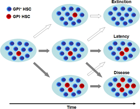
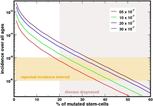
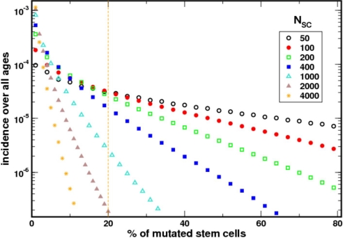
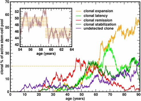
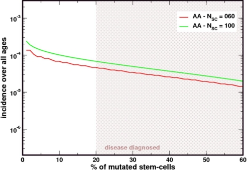
Paroxysmal nocturnal hemoglobinuria is an acquired hematopoietic stem cell (HSC) disorder characterized by the partial or complete deficiency of glycosyl-phosphatidylinositol (GPI)-linked membrane proteins, which leads to intravascular hemolysis. A loss of function mutation in the PIG-A gene, required for GPI biosynthesis, explains how the deficiency of many membrane proteins can result from a single genetic event. However, to date the mechanism of expansion of the GPI(-) clone has not been fully understood. Two hypotheses have been proposed: A selective advantage of GPI(-) cells because of a second mutation or a conditional growth advantage of GPI(-) cells in the presence of an immune attack on normal (GPI(+)) HSCs. Here, we explore a third possibility, whereby the PNH clone does not have a selective advantage. Simulations in a large virtual population accurately reproduce the known incidence of the disease; and the fit is optimized when the number of stem cells is decreased, reflecting a component of bone marrow failure in PNH. The model also accounts for the occurrence of spontaneous cure in PNH, consequent on clonal extinction. Thus, a clonal advantage may not be always necessary to explain clonal expansion in PNH.


Conflict of interest statement

The authors declare no conflict of interest.

Figures

Fig. 1.
Fig. 1.
Model of stochastic dynamics in the active hematopoietic SC pool (NSC). SCs divide at a rate approximately 1/year and acquire a mutation in the PIG-A gene at a rate of 5 × 10⁻⁷ per replication. Cells are selected for replication and export at random. The dynamics of the mutant population (GPI HSCs, red cells) is followed forward in time. Several scenarios become possible because of the stochastic nature of the problem associated with the neutrality of the PIG-A mutation: (Top) Stochastic extinction of the clone, which may be appreciable given the small size of the active HSC pool. (Middle) Latency, in which the PNH burden in the HSC pool is <20%, a situation which can remain as such for several years (see Fig. 4). (Bottom) Disease, in which case the PNH burden in the HSC pool is >20%. The cross-arrows point out to the fact that the situation of a single patient may change from these different stages during their life history, where dark arrows mean the patient condition worsens, whereas light arrows point to changes where patient condition improves.
Fig. 2.
Fig. 2.
Prevalence of PNH based on the US population census. The reported prevalence is 1–10/million population (orange shaded area). In our model, diagnosis requires that ≥20% of the active SCs have a mutation in PIG-A. In an adult this will be ≈80 SCs (and less in a growing child). Our estimate falls well within the expected prevalence of the disease. Different curves were obtained for different mutation rates. The average mutation rate (red curve) is compatible with the prevalence of the disease in the U.S.
Fig. 3.
Fig. 3.
The incidence of the disease depends on the number of HSCs that are actively contributing to hematopoiesis. The incidence predicted by the model matches the epidemiological data optimally when NSC ≈ 400. As the size of the HSCs increases, the incidence of the disease decreases. In these simulations, the rate of HSC replication was kept constant at ≈1/year because there is no experimental data relating how the rate of replication of HSCs changes as their population increases.
Fig. 4.
Fig. 4.
Representative life histories of the PIG-A mutant clone. Once the mutation appears in a HSC, the mutated SC may stochastically expand to become the dominant contributor to hematopoiesis (and PNH will be diagnosed), undergo stochastic extinction, or “stabilize” for many years. Given the limited resolution of standard flow cytometry, the size of the clone may appear “stable” for years (Inset).
Fig. 5.
Fig. 5.
Patients with aplastic anemia (AA) have a higher prevalence of PIG-A mutated SCs. As the size of the stem cell pool decreases as in AA, the prevalence of PIG-A mutants increases. The figure compares the results from Fig. 2 with those obtained by considering a population of 100 or 60 SCs that must replicate at a rate 4 and 6 times faster than normal to maintain hematopoiesis. Under stochastic dynamics and neutral evolution, the clone is more likely to increase in size and appear “more often” in patients with AA.

References

    1. Parker C, et al. Diagnosis and management of paroxysmal nocturnal hemoglobinuria. Blood. 2005;106:3699–3709. - PMC - PubMed
    1. Luzzatto L, Bessler M, Rotoli B. Somatic mutations in paroxysmal nocturnal hemoglobinuria: A blessing in disguise? Cell. 1997;88:1–4. - PubMed
    1. Hillmen P, Bessler M, Mason PJ, Watkins WM, Luzzatto L. Specific defect in N-acetylglucosamine incorporation in the biosynthesis of the glycosylphosphatidylinositol anchor in cloned cell lines from patients with paroxysmal nocturnal hemoglobinuria. Proc Natl Acad Sci USA. 1993;90:5272–5276. - PMC - PubMed
    1. Miyata T, et al. Abnormalities of PIG-A transcripts in granulocytes from patients with paroxysmal nocturnal hemoglobinuria. N Engl J Med. 1994;330:249–255. - PubMed
    1. Rotoli B, Luzzatto L. Paroxysmal nocturnal hemoglobinuria. Semin Hematol. 1989;26:201–207. - PubMed

Publication types

Substances

LinkOut - more resources