. 2009 Feb 15;179(4):279-87.
doi: 10.1164/rccm.200802-320OC. Epub 2008 Nov 14.

Cigarette smoke induces cellular senescence via Werner's syndrome protein down-regulation

Affiliations

Cigarette smoke induces cellular senescence via Werner's syndrome protein down-regulation

Toru Nyunoya et al. Am J Respir Crit Care Med. .

Abstract

Rationale: Werner's syndrome is a genetic disorder that causes premature aging due to loss-of-function mutations in a gene encoding a member of the RecQ helicase family. Both Werner's syndrome and cigarette smoking accelerate aging. No studies have examined the effect of cigarette smoke on Werner's syndrome protein.

Objectives: To investigate the role of Werner's syndrome protein in cigarette smoke-induced cellular senescence.

Methods: Cellular senescence and amounts of Werner's syndrome protein were measured in fibroblasts isolated from patients with emphysema and compared with age-matched nonsmokers. The in vitro effects of cigarette smoke on amounts of Werner's syndrome protein, function, and senescence were also evaluated in primary human lung fibroblasts and epithelial cells.

Measurements and main results: Cultured lung fibroblasts isolated from patients with emphysema exhibited a senescent phenotype accompanied by a decrease in Werner's syndrome protein. Cigarette smoke extract decreased Werner's syndrome protein in cultured fibroblasts and epithelial cells. Werner's syndrome protein-deficient fibroblasts were more susceptible to cigarette smoke-induced cellular senescence and cell migration impairment. In contrast, exogenous overexpression of Werner's syndrome protein attenuated the cigarette smoke effects.

Conclusions: Cigarette smoke induces cellular senescence and cell migration impairment via Werner's syndrome protein down-regulation. Rescue of Werner's syndrome protein down-regulation may represent a potential therapeutic target for smoking-related diseases.


Figures

<b>Figure 1.</b>
Figure 1.
Lung fibroblasts isolated from patients with severe emphysema are deficient in Werner's syndrome protein. (A) Primary lung fibroblasts isolated from three patients with severe emphysema and from three age-matched nonsmokers were cultured for 2 days. Immunoblot analysis was performed for WRN protein. Equal loading was determined by stripping the blot and reprobing with antibodies to β-actin. Densitometry is expressed as arbitrary units for each experimental band (see Immunoblotting). Data are expressed as means ± SEM for each group (n = 3). (B and C) Primary lung fibroblasts were cultured at the starting density of 100 × 103/ml, and cell density and senescence-associated β-galactosidase (SA β-Gal) activity were measured on Day 4. Data are expressed as means ± SEM for each group (n = 3).
<b>Figure 2.</b>
Figure 2.
Cigarette smoke down-regulates Werner's syndrome protein. (A) Primary human lung fibroblasts and HFL-1 cells were cultured with or without 1.2% cigarette smoke extract (CSE) for various periods (1, 2, and 4 d). Immunoblot analysis was performed for WRN protein. Equal loading was determined by stripping the blot and reprobing with antibodies to β-actin. Densitometry is expressed as arbitrary units for each experimental band. Immunoblotting data are representative of three experiments. (B) Primary human tracheobronchial epithelial (hTBE) cells were cultured with or without 2% CSE for various periods (1, 2, and 4 d). Immunoblot analysis was performed for WRN protein. Equal loading was determined by stripping the blot and reprobing with antibodies to β-actin. Densitometry is expressed as arbitrary units for each experimental band. Immunoblotting data are representative of three experiments.
<b>Figure 3.</b>
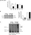
Figure 3.
Cigarette smoke decreases Werner's syndrome protein in a ubiquitin–proteasome-dependent manner. (A) HFL-1 cells were cultured with or without 1.2% CSE. WRN mRNA was measured at various time points (1, 2, and 4 d). Data are expressed as means ± SEM for three independent experiments. Con = control; HPRT = hypoxanthine–guanine phosphoribosyltransferase. (B) HFL-1 cells were cultured with or without 1.5% cigarette smoke extract for 24 hours and then were incubated with 20 μM lactacystin for 3 hours. Immunoblot analysis was performed for WRN protein. Equal loading was determined by stripping the blot and reprobing with antibodies to β-actin. Densitometry is expressed as arbitrary units for each experimental band. Immunoblotting data are representative of three experiments. (C) HFL-1 cells were treated as in (B). After immunoprecipitation of WRN protein, immunoblot analysis was performed for higher molecular weight (HMW) forms of WRN protein. Immunoblotting data are representative of three experiments. NS = nonspecific band.
<b>Figure 4.</b>
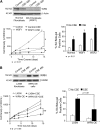
Figure 4.
Modulation of Werner's syndrome protein affects cigarette smoke–induced growth inhibition. (A) WSF1 cells (AG03141) and normal fibroblasts were cultured in the presence or absence of 1.5% cigarette smoke extract (CSE). Immunoblot analysis was performed for WRN protein at 48 hours. Equal loading was determined by stripping the blot and reprobing with antibodies to β-actin. Immunoblotting data are representative of three experiments. The cell counts were also monitored in the presence or absence of 1.2% CSE at various time points (2, 4, 8, and 14 d). SA β-Gal staining was performed for WSF1 cells (AG03141), WSF2 cells (AG12799), and normal fibroblasts in the presence or absence of 1.2% CSE and digital photographs were obtained at 7 days. The percentage of SA β-Gal–positive cells per total cell number is shown. Data are expressed as means ± SEM for three independent experiments (*P < 0.01). (B) HFL-1 cells were transduced with LXSN retroviral vectors encoding complete WRN cDNA. Transduced cells were selected with G418. HFL-1 cells transduced with LXSN-WRN were cultured in the presence or absence of 1.5% CSE. Immunoblot analysis was performed for WRN protein at 48 hours. Equal loading was determined by stripping the blot and reprobing with antibodies to β-actin. Immunoblotting data are representative of three experiments. The cell counts were monitored as in (A). SA β-Gal staining was also performed and digital photographs were obtained at 14 days. The percentage of SA β-Gal–positive cells per total cell number is shown. Data are expressed as means ± SEM for three independent experiments (**P < 0.05). (C) HFL-1 cells transduced with LXSN-WRN were cultured in the presence or absence of 1.5% CSE for 1, 3, and 6 hours. Immunoblot analysis was performed for phosphorylation of histone H2AX. Equal loading was determined by stripping the blot and reprobing with antibodies to β-actin. Densitometry is expressed as arbitrary units for each experimental band. Immunoblotting data are representative of three experiments. WRN OE = WRN protein overexpression.
<b>Figure 5.</b>
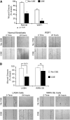
Figure 5.
Modulation of Werner's syndrome protein affects cigarette smoke–induced cell migration impairment. (A) WSF1 cells (AG03141) or normal fibroblasts were cultured until confluence in 12-well tissue culture plates and were further incubated with or without 1.5% cigarette smoke extract (CSE). After creating a scratch wound, the medium was refreshed with or without 1.5% CSE. Closure of the open wound area at 24 hours is shown as a percentage compared with the area of the initial wound. (B) HFL-1 cells transduced with LXSN-WRN were treated as in (A). Data are expressed as means ± SEM for three independent experiments (*P < 0.01). Digital photographs were obtained at 0 and 24 hours.
<b>Figure 6.</b>
Figure 6.
N-Acetylcysteine (NAC) alleviates Werner's syndrome protein down-regulation and growth inhibition in cigarette smoke–exposed fibroblasts. (A) WSF1 cells (AG03141) in the absence of cigarette smoke extract (CSE) and normal dermal fibroblasts in the absence or presence of CSE were cultured for 48 hours, and then incubated at 37°C for 40 minutes with dihydroethidium (DHE, 10 μmol/L) in 2 ml of phosphate-buffered saline containing pyruvate (5 mmol/L). Cells were trypsinized on ice and analyzed by flow cytometry. The mean fluorescence intensity (MFI) of each sample (10,000 cells) was measured. Values indicate the ratio of MFI relative to control MFI. Data are expressed as means ± SEM for three independent experiments. (B) HFL-1 cells were cultured in the presence or absence of 40 μM acrolein, or 200 μM hydrogen peroxide, for 24 hours. Western blot analysis was performed for WRN protein. Equal loading was determined by stripping the blot and reprobing with antibodies to β-actin. Immunoblotting data are representative of three experiments. (C) human pulmonary fibroblasts (HPFs) were cultured with or without 1.5% CSE in the presence or absence of 3 mM NAC for 48 hours. Immunoblot analysis was performed for WRN protein. Equal loading was determined by stripping the blot and reprobing with antibodies to β-actin. Immunoblotting data are representative of three experiments. (D) HPFs were treated as in (A). Cell density was measured at 48 hours. Data are expressed as means ± SEM for three independent experiments.

References

    1. Martin GM. Genetic syndromes in man with potential relevance to the pathobiology of aging. Birth Defects Orig Artic Ser 1978;14:5–39. - PubMed
    1. Epstein CJ, Martin GM, Schultz AL, Motulsky AG. Werner's syndrome: a review of its symptomatology, natural history, pathologic features, genetics and relationship to the natural aging process. Medicine (Baltimore) 1966;45:177–221. - PubMed
    1. Goto M, Tanimoto K, Horiuchi Y, Sasazuki T. Family analysis of Werner's syndrome: a survey of 42 Japanese families with a review of the literature. Clin Genet 1981;19:8–15. - PubMed
    1. Brown WT, Kieras FJ, Houck GE Jr, Dutkowski R, Jenkins EC. A comparison of adult and childhood progerias: Werner syndrome and Hutchinson-Gilford progeria syndrome. Adv Exp Med Biol 1985;190:229–244. - PubMed
    1. Martin GM, Oshima J, Gray MD, Poot M. What geriatricians should know about the Werner syndrome. J Am Geriatr Soc 1999;47:1136–1144. - PubMed

Publication types