Comparative Study
. 2009 Feb 1;179(3):212-9.
doi: 10.1164/rccm.200802-228OC. Epub 2008 Nov 14.

Insulin-like growth factor-I receptor blockade improves outcome in mouse model of lung injury

Affiliations
Comparative Study

Insulin-like growth factor-I receptor blockade improves outcome in mouse model of lung injury

Jung-Eun Choi et al. Am J Respir Crit Care Med. .

Abstract

Rationale: The insulin-like growth factor-I (IGF-I) pathway is an important determinant of survival and proliferation in many cells. However, little is known about the role of the IGF-I pathway in lung injury. We previously showed elevated levels of IGF-I in bronchoalveolar lavage fluid from patients with acute respiratory distress syndrome. Furthermore, immunodepletion of IGF from acute respiratory distress syndrome bronchoalveolar lavage increased fibroblast apoptosis.

Objectives: We examined the effect of blockade of type 1 IGF tyrosine kinase receptor (IGF-IR) in a murine model of bleomycin-induced lung injury and fibrosis.

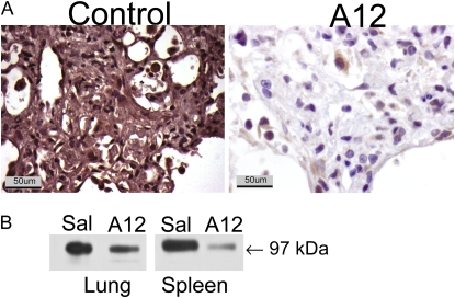
Methods: Mice were treated with a monoclonal antibody against the IGF-I receptor (A12) or vehicle after intratracheal bleomycin instillation.

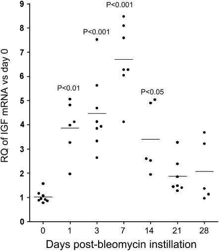
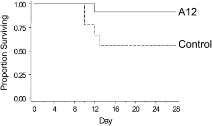
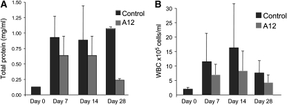
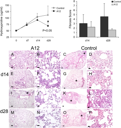
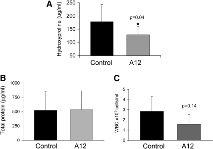
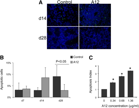
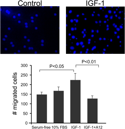
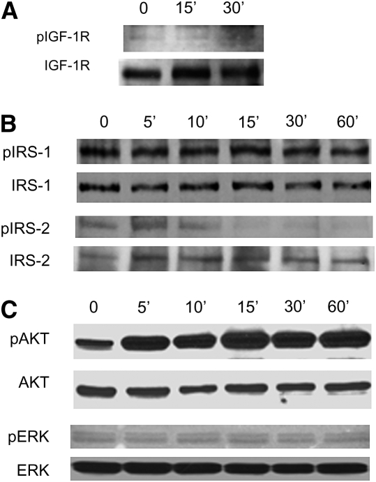
Measurements and main results: Mice treated with A12 antibody had significantly improved survival after bleomycin injury compared with control mice. Both groups of mice had a similar degree of fibrosis on days 7 and 14, but by Day 28 the A12-treated group had significantly less fibrosis. Delayed treatment with A12 also resulted in decreased fibrosis. A12-treated mice had significantly decreased apoptotic cells on Day 28 compared with control mice. We confirmed that A12 treatment induced mouse lung fibroblast apoptosis in vitro. In addition, IGF-I increased lung fibroblast migration. The primary pathway activated by IGF-I in lung fibroblasts was the insulin receptor substrate-2/phosphatidylinositol 3-kinase/Akt axis.

Conclusions: IGF-I regulated survival and migration of fibrogenic cells in the lung. Blockade of the IGF pathway increased fibroblast apoptosis and subsequent resolution of pulmonary fibrosis. Thus, IGF-IR may be a potential target for treatment of lung injury and fibrosis.


Figures

<b>Figure 1.</b>
Figure 1.
Real-time polymerase chain reaction analysis of insulin-like growth factor (IGF) mRNA expression after bleomycin administration. Data are normalized to hypoxanthine–guanine phosphoribosyltransferase expression. The y axis represents fold increase compared with Day 0. Each point represents an individual mouse. Mean values are indicated. RQ = relative quantity.
<b>Figure 2.</b>
Figure 2.
Decreased insulin-like growth factor-I receptor (IGF-IR) expression after systemic administration of A12, a monoclonal antibody against IGF-IR. (A) Immunohistochemical analysis for IGF-IR was performed on lungs on Day 7 after bleomycin administration. (B) Western blot analysis for IGF-IR, performed on lung and spleen lysates on Day 14 after bleomycin administration to mice treated with saline (Sal) or A12.
<b>Figure 3.</b>
Figure 3.
Kaplan-Meier survival curves for control and A12-treated mice after bleomycin treatment. The difference in survival was significant: P = 0.038, A12 versus saline (control).
<b>Figure 4.</b>
Figure 4.
Bronchoalveolar lavage fluid (BALF) total protein concentrations (A) and cell count (B) at 0 (baseline), 7, 14, and 28 days after bleomycin instillation in A12-treated mice and control mice. Average values and SD are shown. WBC = white blood cells.
<b>Figure 5.</b>
Figure 5.
Top: Hydroxyproline content (left) and fibrosis scores (right) from A12-treated mice and control mice after bleomycin instillation. Data were analyzed by two-way analysis of variance (ANOVA) with Tukey's honestly significant difference (HSD) post-hoc test, and statistical significance was set at *P < 0.05. Bottom: Histology of A12-treated mice and control mice at 14 days (AH) and 28 days (IP) after bleomycin instillation. Lung sections from A12-treated mice show relatively normal interstitium and less fibrosis (*) compared with control mice. Right middle lobe samples from two mice per group are shown. Original magnification: (A, C, E, G, I, K, M, and O) ×4; (B, D, F, H, J, L, N, and P) ×40. Scale bars: (A, C, E, G, I, K, M, and O) 200 μm; (B, D, F, H, J, L, N, and P) 25 μm. Stained with hematoxylin and eosin.
<b>Figure 6.</b>
Figure 6.
Hydroxyproline content (A), bronchoalveolar lavage fluid (BALF) total protein (B), and BALF total cell count (C) on Day 21 after bleomycin instillation and initiation of A12 antibody administration on Day 7. n = at least five animals per group. Average values and SD are shown.
<b>Figure 7.</b>
Figure 7.
(A) Terminal deoxynucleotidyltransferase dUTP nick end labeling (TUNEL)-positive cells (green) in saline- and A12-treated mouse lungs on Days 14 and 28 after bleomycin injury. Nuclei are counterstained with 4′,6-diamidino-2-phenylindole (DAPI). Original magnification: ×20. (B) Quantification of TUNEL IHC by determining the number of TUNEL-positive cells per total number of cells. Shown is the average and SD of two mice per condition, with at least 500 cells analyzed per condition. Data were analyzed by two-way analysis of variance with Tukey's honestly significant difference (HSD) post-hoc test. (C) Increased apoptosis of primary mouse lung fibroblasts treated with insulin-like growth factor-I receptor (IGF-IR) antibody. Mouse lung fibroblasts were serum starved overnight and then incubated with the indicated concentration of IGF-IR antibody A12. Apoptosis was measured with Cell Death Detection ELISAPLUS (Roche Applied Science). All experiments were done in triplicate and repeated at least twice. The apoptosis index is defined as follows: the ratio of the optical density at 405 nm (OD405 nm) of the experimental condition to that of the control condition (medium alone). *P < 0.05 compared with the control group.
<b>Figure 8.</b>
Figure 8.
Fibroblast proliferation in response to insulin-like growth factor (IGF). Mouse lung fibroblasts were plated in triplicate and serum starved overnight followed by addition of IGF-I (100 ng/ml), serum-free medium (negative control), or 10% fetal bovine serum (FBS; positive control) and then incubated for the indicated times. Cell proliferation was measured by cell proliferation assay (MTT; Roche, Indianapolis, IN) assay. Data are shown as mean OD570 nm and SD and represent the average of at least four independent experiments.
<b>Figure 9.</b>
Figure 9.
Insulin-like growth factor-I (IGF-I)–induced migration of fibroblasts. Mouse lung fibroblasts were plated on FluoroBlok transwell filters overnight, and then IGF-I (100 ng/ml), serum-free medium (negative control), or 10% serum (positive control) was added to the lower chamber. Some cells were preincubated with the blocking antibody to IGF-I receptor (A12, 40 μg/ml) before the addition of IGF-I. Cells were allowed to migrate through the membrane for 4 hours at 37°C. Migrated cells were counted by fluorescence microscopy. Each experiment was done in triplicate and repeated at least three times. Average cell counts and SD are presented.
<b>Figure 10.</b>
Figure 10.
Mouse lung fibroblasts were serum starved overnight and then stimulated with insulin-like growth factor-I (100 ng/ml) for the indicated times. Cells were lysed, and proteins were separated by sodium dodecyl sulfate–polyacrylamide gel electrophoresis. Each membrane was blotted with the indicated phospho-antibody [(A) IGF-1R; (B) IRS-1, IRS-2 (C) AKT, ERK] and then stripped and reblotted with antibody to total protein.

References

    1. LeRoith D, Roberts CT Jr. The insulin-like growth factor system and cancer. Cancer Lett 2003;195:127–137. - PubMed
    1. Kurmasheva RT, Houghton PJ. IGF-I mediated survival pathways in normal and malignant cells. Biochim Biophys Acta 2006;1766:1–22. - PubMed
    1. Rom WN, Basset P, Fells GA, Nukiwa T, Trapnell BC, Crysal RG. Alveolar macrophages release an insulin-like growth factor I–type molecule. J Clin Invest 1988;82:1685–1693. - PMC - PubMed
    1. Maeda A, Hiyama K, Yamakido H, Ishioka S, Yamakido M. Increased expression of platelet-derived growth factor A and insulin-like growth factor-I in BAL cells during the development of bleomycin-induced pulmonary fibrosis in mice. Chest 1996;109:780–786. - PubMed
    1. Krein PM, Sabatini PJ, Tinmouth W, Green FH, Winston BW. Localization of insulin-like growth factor-I in lung tissues of patients with fibroproliferative acute respiratory distress syndrome. Am J Respir Crit Care Med 2003;167:83–90. - PubMed

Publication types

MeSH terms