. 2008 Dec 18;456(7224):989-92.
doi: 10.1038/nature07468. Epub 2008 Nov 16.

Structural recognition and functional activation of FcgammaR by innate pentraxins

Affiliations

Structural recognition and functional activation of FcgammaR by innate pentraxins

Jinghua Lu et al. Nature. .

Abstract

Pentraxins are a family of ancient innate immune mediators conserved throughout evolution. The classical pentraxins include serum amyloid P component (SAP) and C-reactive protein, which are two of the acute-phase proteins synthesized in response to infection. Both recognize microbial pathogens and activate the classical complement pathway through C1q (refs 3 and 4). More recently, members of the pentraxin family were found to interact with cell-surface Fcgamma receptors (FcgammaR) and activate leukocyte-mediated phagocytosis. Here we describe the structural mechanism for pentraxin's binding to FcgammaR and its functional activation of FcgammaR-mediated phagocytosis and cytokine secretion. The complex structure between human SAP and FcgammaRIIa reveals a diagonally bound receptor on each SAP pentamer with both D1 and D2 domains of the receptor contacting the ridge helices from two SAP subunits. The 1:1 stoichiometry between SAP and FcgammaRIIa infers the requirement for multivalent pathogen binding for receptor aggregation. Mutational and binding studies show that pentraxins are diverse in their binding specificity for FcgammaR isoforms but conserved in their recognition structure. The shared binding site for SAP and IgG results in competition for FcgammaR binding and the inhibition of immune-complex-mediated phagocytosis by soluble pentraxins. These results establish antibody-like functions for pentraxins in the FcgammaR pathway, suggest an evolutionary overlap between the innate and adaptive immune systems, and have new therapeutic implications for autoimmune diseases.


Figures
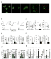
Figure 1
Figure 1
Pentraxin activation of FcγR results in opsonization and cytokine release. a) SAP- and CRP-opsonized zymosan are phagocytosed by human macrophages through FcγRIIa. MDM incubated with zymosan (red) opsonized with (from left to right) SAP, CRP, IgG, PBS control, or SAP in the presence of 10 mg/ml IVIg. b-f) The production of IL-10, IL-8 and IL-6 by CD14+ monocytes in response to b) different concentrations of aggregated SAP; c) 50 μg/ml aggregated or monomeric SAP; d) SAP pre-treated with proteinase K or pre-cleared with PE-Sepharose; and e) SAP in the presence of 25 μg/ml piceatannol; f) SAP treatment in the presence of FcγR specific antibodies or control Ig. g) Cytokine release by BMDM from wild type, MyD88-/- and RIP2-/- mice stimulated with SAP.
Figure 2
Figure 2
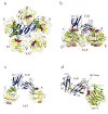
Crystal structure of SAP-FcγRIIa complex. The view is from the face (a) and side (b and c) of SAP, with panel c highlighting only the receptor contact A and C subunits. The five SAP subunits are shown in yellow with ridge helices in red, and FcγRIIa is colored in blue. The interface is represented by molecular surface in green. The calcium and ligand binding sites on SAP are highlighted in magenta. d) Comparison between the free (green) and receptor-bound (yellow) SAP, and between the free (wheat) and SAP-bound (blue) FcγRIIa structures. For clarity, only the A subunit is shown from the superposition of SAP pentamer. The BC loop (residues 28-35) of D1 domain is indicated.
Figure 3
Figure 3
The binding interfaces between SAP and FcγRIIa. a) The interface between the D1 domain of FcγRIIa (blue and magenta) and the A subunit of SAP (yellow and green) is shown with participating side residues shown in sticks. The hydrogen bond interactions are represented by red dashed lines. b) The interface between the D2 domain of FcγRIIa and the C subunit of SAP.
Figure 4
Figure 4
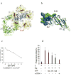
Competition between human IgG1 and SAP or CRP for binding to Fcγ receptors. a) The superposition of FcγR between SAP-FcγRIIa and Fc-FcγRIII complexes with FcγRIIa, Fc portion of IgG1 and SAP shown in blue, green and yellow, respectively. b) The interface residues of SAP-FcγRIIa and Fc-FcγRIII complexes are depicted by molecular surface representations in blue and green on FcγRIIa (blue) and FcγRIII (green), respectively. c and e) Competition binding between SAP (c), CRP (e) and human IgG1 using SAP or CRP immobilized CM5 sensorchips. The analytes consisted of a mixture of (c) 5 μM and (e) 2 μM of stated FcγR with various concentrations of hIgG1. d) SAP and f) CRP inhibit IgG-mediated phagocytosis. Human MDM incubated with E or EIg, in the presence of various concentrations (μg/ml) of CRP, SAP, or blocking antibodies against FcγRI (25 μg/ml) and FcγRII (25μg/ml).
Figure 4
Figure 4
Competition between human IgG1 and SAP or CRP for binding to Fcγ receptors. a) The superposition of FcγR between SAP-FcγRIIa and Fc-FcγRIII complexes with FcγRIIa, Fc portion of IgG1 and SAP shown in blue, green and yellow, respectively. b) The interface residues of SAP-FcγRIIa and Fc-FcγRIII complexes are depicted by molecular surface representations in blue and green on FcγRIIa (blue) and FcγRIII (green), respectively. c and e) Competition binding between SAP (c), CRP (e) and human IgG1 using SAP or CRP immobilized CM5 sensorchips. The analytes consisted of a mixture of (c) 5 μM and (e) 2 μM of stated FcγR with various concentrations of hIgG1. d) SAP and f) CRP inhibit IgG-mediated phagocytosis. Human MDM incubated with E or EIg, in the presence of various concentrations (μg/ml) of CRP, SAP, or blocking antibodies against FcγRI (25 μg/ml) and FcγRII (25μg/ml).

References

    1. Steel DM, Whitehead AS. The major acute phase reactants: C-reactive protein, serum amyloid P component and serum amyloid A protein. Immunol Today. 1994;15:81–88. - PubMed
    1. Pepys MB, et al. Comparative clinical study of protein SAP (amyloid P component) and C-reactive protein in serum. Clin Exp Immunol. 1978;32:119–124. - PMC - PubMed
    1. Garlanda C, Bottazzi B, Bastone A, Mantovani A. Pentraxins at the crossroads between innate immunity, inflammation, matrix deposition, and female fertility. Annu Rev Immunol. 2005;23:337–366. - PubMed
    1. Kaplan MH, Volanakis JE. Interaction of C-reactive protein complexes with the complement system. I. Consumption of human complement associated with the reaction of C-reactive protein with pneumococcal C-polysaccharide and with the choline phosphatides, lecithin and sphingomyelin. J Immunol. 1974;112:2135–2147. - PubMed
    1. Bharadwaj D, Stein MP, Volzer M, Mold C, Du Clos TW. The major receptor for C-reactive protein on leukocytes is fcgamma receptor II. J Exp Med. 1999;190:585–590. - PMC - PubMed

Publication types

MeSH terms

Associated data