. 2008 Oct;12(5B):2062-72.
doi: 10.1111/j.1582-4934.2008.00199.x.

Neovascularization in an arterio-venous loop-containing tissue engineering chamber: role of NADPH oxidase

Affiliations

Neovascularization in an arterio-venous loop-containing tissue engineering chamber: role of NADPH oxidase

F Jiang et al. J Cell Mol Med. 2008 Oct.

Abstract

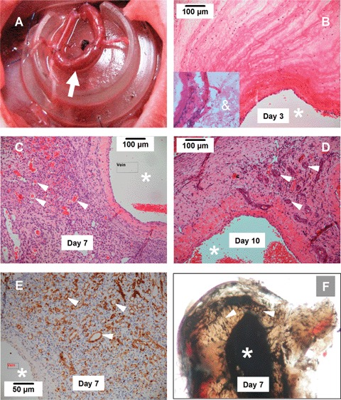
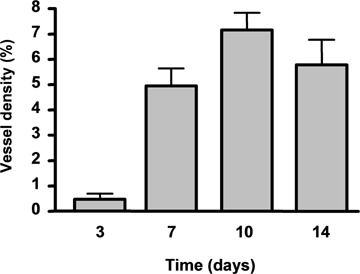
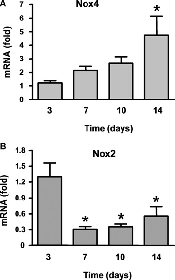
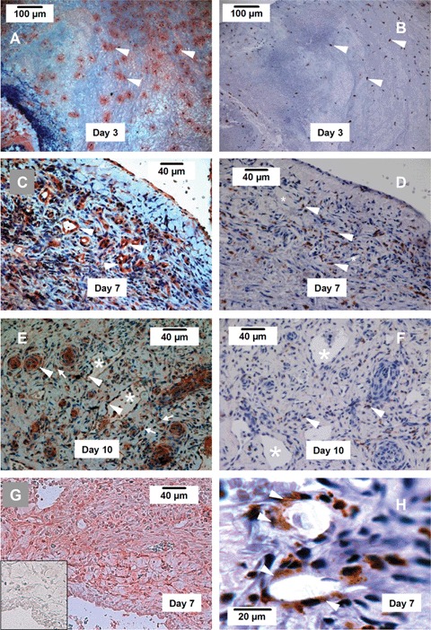
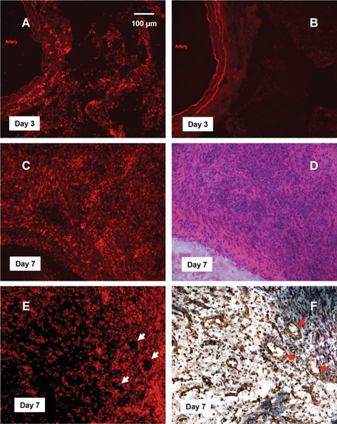
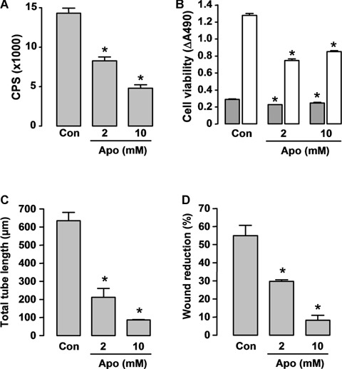
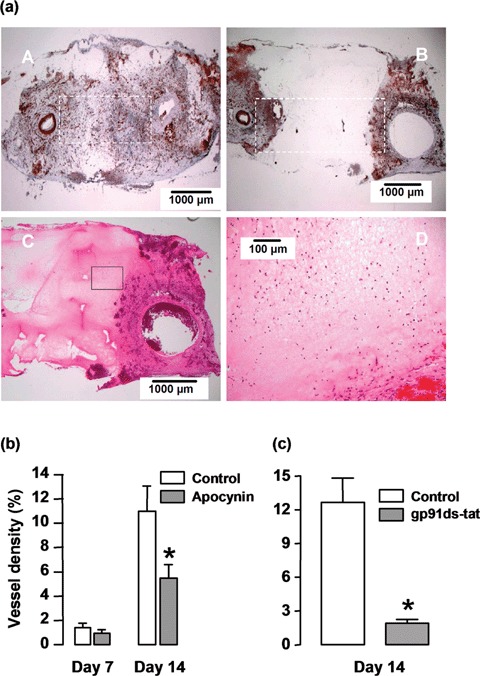
Using an in vivo arterio-venous loop-containing tissue-engineering chamber, we have created a variety of vascularized tissue blocks, including functional myocardium. The viability of the transplanted cells is limited by the rate of neovascularization in the chamber. A Nox2-containing nicotinamide adenine dinucleotide phosphate (NADPH) oxidase is thought to have a critical role in ischaemic angiogenesis. In this study we investigated whether NADPH oxidase is involved in the neovascularization process in the tissue-engineering chamber. New blood vessels originating from the venous and the arterial ends of the loop could be identified after 3 days, and the vessel density (by lectin staining) peaked after 7 days and was maintained for at least 14 days. This was accompanied by granulation tissue formation and concomitant increase in the mRNA level of Nox4 NADPH oxidase. Although the total level of Nox2 mRNA in the chamber tissue decreased from day 3 to day 7, immunohistochemistry identified a strong expression of Nox2 in the endothelial cells of the new vessels. In human microvascular endothelial cells, the NADPH oxidase inhibitor apocynin reduced NADPH oxidase activity and inhibited the angiogenic responses in vitro. Local treatment with the NADPH oxidase inhibitors apocynin or gp91ds-tat peptide significantly suppressed the vessel growth in the chamber. In conclusion, NADPH oxidase-dependent redox signalling is important for neovascularization in this novel tissue-engineering chamber in vivo, and boosting this signalling might be a new approach to extending vascularization and tissue growth.


Figures

Fig 1
Fig 1
Angiogenic response in tissue-engineering (TE) chamber (A). Arrow indicates the arterio-venous loop constructed by a vein graft anastomosed to the femoral artery and vein. (B-D) Histology of the tissue formed in the TE chamber 3, 7 and 10 days after chamber implantation (H&E staining). The lumen of the vein side of the parent loop was indicated by *. The new blood vessels are indicated by arrow heads. Note that vessels with a multi-layered wall composed of mural cells can be observed in 10-day specimen. The insert in B shows that 1 day after construction, the chamber contained only a fibrin mesh (&) and a few infiltrating cells. (E) New vessels defined by Griffonia Simplicifolia lectin staining (brown, arrow heads) at 7 days. (F) Ink perfusion of the AV loop at 7 days demonstrates new vessels (arrow heads) sprouting form the parent vessel lumen (femoral vein). At each time point, 5–6 animals were analysed.
Fig 2
Fig 2
Density (% area) of new vessels in the chamber tissue 3,7,10 and 14 days after chamber implantation. Data are mean ± standard error of the mean (SEM), n = 3–4.
Fig 3
Fig 3
Time course of the expression levels of mRNA of the NADPH oxidase Nox4 and Nox2 in the chamber tissue. Data (mean ± S.E.M.) are expressed as fold of the mean level of day 3. *P < 0.05 versus day 3, one-way analysis of the variance (anova) followed by Newman-Keuls t-test, n = 6–7.
Fig 4
Fig 4
Immunohistochemistry showing the expression of Nox2, Nox4 and the mono-cytes/macrophage marker ED-1. (A, C, E) Nox2 expression (red) in new blood vessels (arrow heads) and infiltrating leukocytes (arrows) in the chamber tissue at 3, 7 and 10 days. (B, D, F) Expression of ED-1 (brown) in infiltrating leukocytes (arrow heads) in the chamber tissue at 3, 7 and 10 days. (G) Expression of Nox4 (red colour) in the chamber tissue at 7 days. The insert shows a negative control section, in which non-specific IgG was used instead of the primary antibody. (H) We have observed that at day 7, some ED-1 + cells appeared to be incorporated in the new vessel (arrow heads). Such ED-1 + cells could not be observed in the vessels at day 10. Endothelial cells of the new vessel were ED-1 negative. * indicates the lumen of new blood vessels, n = 4 at each time point.
Fig 5
Fig 5
In situ superoxide production detected by dihydroethidium (DHE) fluorescence. (A, C) superoxide production in 3- and 7-day specimens respectively. (B) A 3-day sample pre-incubated with the superoxide dismutase mimetic Mn(lll)TMPyP, showing that the fluorescence was superoxide specific. Some autofluorescence was observed only in the elastic layers of the parent AV loop. (D) H&E staining of the 7-day sample, showing that intense DHE fluorescence was present in the highly cellularized granulation tissue with numerous new vessels. (E, F) Comparison of DHE fluorescence with lectin reactivity in consecutive sections demonstrated that some of the fluorescent cells formed ring structures that resembled the new blood vessels (arrows), n = 3 at each time point.
Fig 6
Fig 6
Effects of the nicotinamide adenine dinucleotide phosphate (NADPH) oxidase inhibitor apocynin on (A) NADPH oxidase activity, (B) cell viability and (C, D) angiogenic responses in cultured Human microvascular endothelial cells (HMEC) cells. (A) Cells were detached by trypsinization and resuspended in Hank's balanced salt solution. Apocynin (Apo) was added 60 min before assay. Superoxide release was measured by lucigenin (5 μM)-enhanced chemiluminescence after stimulation with 100 μM NADPH in a topcount microplate reader (model 9912, Packard). The results are expressed as count per second (CPS) per 105 cells (n = 5). (C) Con, DMSO control. (B) Cell survival was assessed 24 (grey bars) and 72 hrs (hatched bars) after apocynin treatment with the CellTiter-96 AQueous One Solution kit, and the results are expressed as differences of absorbance at 490 nm between wells containing cells and buffer only (background) (n = 4). (C) In vitro angiogenesis was assessed by the tube formation assay (observed 6 hrs after cell plating on Matrigel, n = 3). Apocynin was added at the time of plating. (D) Wound-healing assay in HMECs observed 18 hrs after scratching (n = 3). Apocynin was added at the time of scratching. * P < 0.05 versus corresponding control (Con), one-way anova.
Fig 7
Fig 7
Effects of NADPH oxidase inhibitors apocynin and gp91ds-tat on angiogenesis in the chamber in vivo. (A, panels A, B) lectin staining of sections from the 14-day samples without (panel A) and with (panel B) apocynin (10 mM) treatment, (panel C) H&E staining showing that the apocynin-containing Matrigel (the pink matrix) did not prevent the infiltration of inflammatory cells prior to vessel development in unorganized areas, (panel D) higher magnification of the outlined area in panel C. (b) Quantitative data of the vessel density (% area) in control and apocynin-treated chamber tissues at 7 and 14 days. The vessel density was assessed in an area as outlined in panels A & B. * P<0.05 versus control, unpaired t-test, n = 5. (c) Effects of gp91ds-tat (100 μM) on neovascularization. A scrambled peptide was used as control. * P < 0.005 versus control, unpaired t-test, n = 5.

References

    1. Cassell OC. Hofer SO, Morrison WA, Knight KR. Vascularisation of tissue-engineered grafts: the regulation of angiogenesis in reconstructive surgery and in disease states. Br J Plast Surg. 2002;55:603–10. - PubMed
    1. Cassell OC, Morrison WA, Messina A, Penington AJ, Thompson EW, Stevens GW, Perera JM, Kleinman HK, Hurley JV, Romeo R, Knight KR. The influence of extracellular matrix on the generation of vascularized, engineered, transplantable tissue. Ann N YAcad Sci. 2001;944:429–42. - PubMed
    1. Hofer SO, Knight KM, Cooper-White JJ, O’Connor AJ, Perera JM, Romeo-Meeuw R, Penington AJ, Knight KR, Morrison WA, Messina A. Increasing the volume of vascularized tissue formation in engineered constructs: an experimental study in rats. Plast Reconstr Surg. 2003;111:1186–92. ; discussion 93–4. - PubMed
    1. Messina A, Bortolotto SK, Cassell OC, Kelly J, Abberton KM, Morrison WA. Generation of a vascularized organoid using skeletal muscle as the inductive source. FASEB J. 2005;19:1570–2. - PubMed
    1. Mian R, Morrison WA, Hurley JV, Penington AJ, Romeo R, Tanaka Y, Knight KR. Formation of new tissue from an arte-riovenous loop in the absence of added extracellular matrix. Tissue Eng. 2000;6:595–603. - PubMed

Publication types

MeSH terms