. 2009 Jan;108(1):285-94.
doi: 10.1111/j.1471-4159.2008.05765.x. Epub 2008 Nov 19.

Developmental and daily expression of the Pax4 and Pax6 homeobox genes in the rat retina: localization of Pax4 in photoreceptor cells

Affiliations

Developmental and daily expression of the Pax4 and Pax6 homeobox genes in the rat retina: localization of Pax4 in photoreceptor cells

Martin F Rath et al. J Neurochem. 2009 Jan.

Abstract

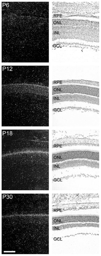
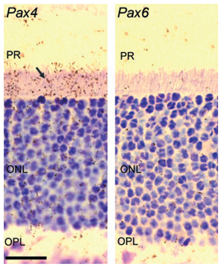
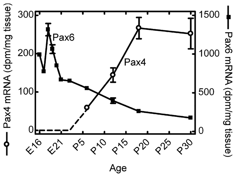
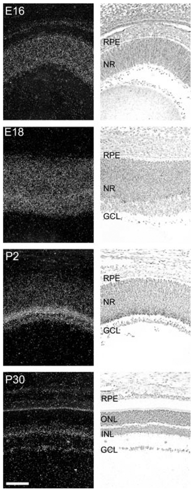
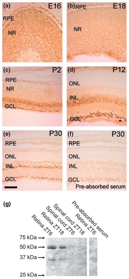
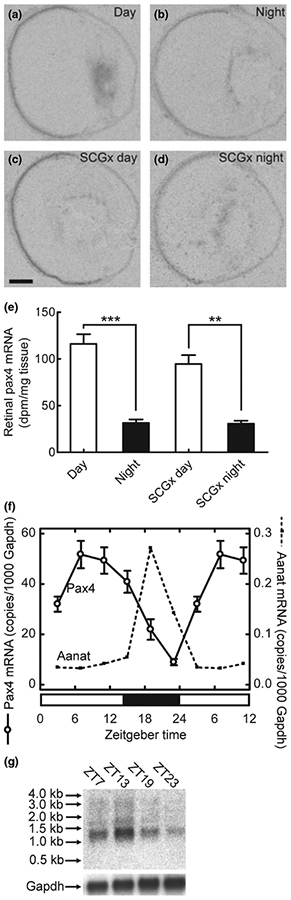
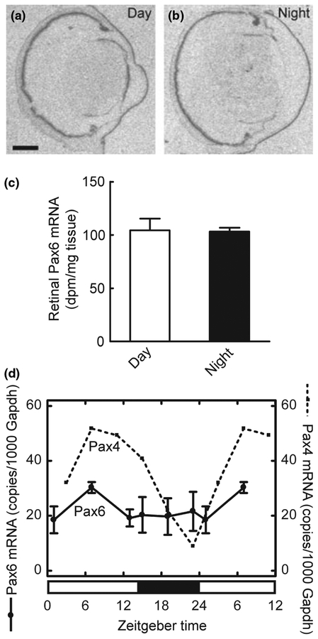
Pax4 is a homeobox gene encoding Pax4, a transcription factor that is essential for embryonic development of the endocrine pancreas. In the pancreas, Pax4 counters the effects of the related transcription factor, Pax6, which is known to be essential for eye morphogenesis. In this study, we have discovered that Pax4 is strongly expressed in retinal photoreceptors of the rat. Pax4 expression is not detectable in the foetal eye; however, postnatal Pax4 transcript levels rapidly increase. In contrast, Pax6 exhibits an inverse developmental pattern of expression being more strongly expressed in the foetal eye. Histological analysis revealed that Pax4 mRNA is exclusively expressed in the retinal photoreceptors, whereas Pax6 mRNA and protein are present in the inner nuclear layer and in the ganglion cell layer of the mature retina. In the adult retina, Pax4 transcripts exhibit a diurnal rhythm with maximal levels occurring during the light period, whereas retinal Pax6 transcript levels do not change throughout the day. The daily changes in Pax4 expression may contribute to daily changes in function in the differentiated retinal photoreceptor.


Figures

Fig. 1
Fig. 1
Ontogenetic expression of Pax4 in the rat retina. Radiochemical in situ hybridization for detection of Pax4 mRNA in sections of the rat retina. The last four stages, postnatal day 6 (P6), P12, P18 and P30, of a developmental series ranging from embryonic day 16 (E16) to P30 are presented. Left column: dark field photomicrographs of hybridized sections dipped in a photographic emulsion. Right column: bright field photomicrographs of the same hybridized sections. Photomicrographs of a section hybridized with a sense control probe are provided in Fig. S1. GCL, ganglion cell layer; INL, inner nuclear layer; ONL, outer nuclear layer; RPE, retinal pigment epithelium. Scale bar, 100 μm.
Fig. 2
Fig. 2
Radiochemical in situ hybridization for detection of Pax4 (left) and Pax6 mRNA (right) in the outer part of the neural retina of the rat at postnatal day 30 (P30). The arrow indicates the positive hybridization signal in the inner segments of the photoreceptors in the section hybridized for detection of Pax4 mRNA. Photomicrographs of a section hybridized with a sense control probe are provided in Fig. S1. Pax6 mRNA is not detected in the photoreceptor cells at P30. ONL, outer nuclear layer; OPL, outer plexiform layer; PR, photoreceptors. Scale bar, 20 μm.
Fig. 3
Fig. 3
Densitometric quantification of in situ hybridization autoradiographs of ontogenetic Pax4 and Pax6 gene expression in the rat retina. In the earliest stages, a Pax4 signal above background was not detected (dotted line). Values on graphs represent the mean ± SEM of three animals at each developmental stage examined. Significant differences in Pax4 mRNA levels occurred during development (one-way ANOVA, F3,8 = 14.2, p = 0.0015) with maximal levels detected at postnatal day 18 (P18) and P30 (Tukey’s multiple comparison test, p-values < 0.05). Significant differences in Pax6 mRNA levels during development were also detected (one-way ANOVA, F10,21 = 130.8, p < 0.0001) with maximal levels obtained at embryonic day 18 (E18) (Tukey’s multiple comparison test, p-values < 0.001).
Fig. 4
Fig. 4
Ontogenetic expression of Pax6 in the rat retina. Radiochemical in situ hybridization for detection of Pax6 mRNA in sections of the rat retina. Images of representative stages of a developmental series ranging from embryonic day (E16) to postnatal day (P30) are displayed. Left column: dark field photomicrographs of hybridized sections dipped in a photographic emulsion. The bright line corresponding to the inner segments of the photoreceptors on the P30 image is because of an endogenous optic scattering of light in this layer and does not represent a hybridization signal. Right column: bright field photomicrographs of the same hybridized sections. Photomicrographs of a section hybridized with a sense control probe are provided in Fig. S1. GCL, ganglion cell layer; INL, inner nuclear layer; NR, neural retina; ONL, outer nuclear layer; RPE, retinal pigment epithelium. Scale bar, 100 μm.
Fig. 5
Fig. 5
Pax6 protein in the rat retina. (a–e) Immunohistochemical analysis of Pax6 protein in the developing rat retina. Photomicrographs of representative developmental stages, as indicated in the upper right corner of each photomicrograph, are displayed. Note the strong immunoreactivity in the nuclei of immunoreactive cells. (f) Immunohistochemical pre-absorption control. GCL, ganglion cell layer; INL, inner nuclear layer; NR, neural retina; ONL, outer nuclear layer; RPE, retinal pigment epithelium. Scale bar, 100 μm. (g) Western blot analysis of the presence of Pax6 protein in the retina and spinal cord of adult rats killed at zeitgeber time (ZT6) and ZT18, respectively. The right hand part of the blot displays a pre-absorption control. Rat Pax6 protein (NP_037133) has a predicted molecular weight of 46.8 kDa. Arrows indicate molecular weight markers. P, postnatal day; E, embryonic day.
Fig. 6
Fig. 6
Diurnal expression of Pax4 in the retina of the adult rat. (a–e) Quantitative radiochemical in situ hybridization analysis of a diurnal variation in expression of Pax4 in the retina of the adult rat housed under a 12 : 12 (light : dark) schedule. (a) Autoradiograph of a section of the eyeball from an animal killed at midday zeitgeber time (ZT6). (b) Autoradiograph of a section of the eyeball from an animal killed at midnight (ZT18). (c) Autoradiograph of a section of the eyeball from a superior cervical ganglionectomized animal killed at midday (ZT6). (d) Autoradiograph of a section of the eyeball from a superior cervical ganglionectomized animal killed at midnight (ZT18). (e) Densitometric quantification of Pax4 mRNA in the rat retina. Values on the bar graph represent the mean ± SEM of four animals in each experimental group. Time of sampling was found to significantly influence retinal Pax4 mRNA levels (two-way ANOVA, F1,12 = 102.5, p < 0.0001), whereas an effect of surgery was not detected (two-way ANOVA, F1,12 = 2.3, p = 0.15). Pair-wise comparison revealed a significant difference in retinal Pax4 expression between day and night in intact animals (two-tailed Student’s t-test, t6 = 7.7, p = 0.00025) and in ganglionectomized animals (two-tailed Student’s t-test, t5 = 5.6, p = 0.0024). (f) Quantitative RT-PCR analysis of diurnal expression ofPax4 and Aanat in the retina of adult rats housed under a 14 : 10 light/dark schedule. Nine animals were killed at each of six time points throughout the 24-hour period. Values on graphs represent the mean ± SEM of three different pools of retinae at each time point. Pax4 mRNA levels changed significantly during the 24-hour period (one-way ANOVA, F5,12 = 15.9, p < 0.0001) with transcript levels at ZT7 being higher than levels at ZT19 (two-tailed Student’s t-test, t4 = 4.4, p = 0.012). Aanat mRNA levels changed significantly during the 24-hour period (one-way ANOVA, F5,12 = 33.1, p < 0.0001) with levels at ZT19 being higher than levels at ZT7 (two-tailed Student’s t-test, t4 = 7.2, p = 0.002). (g) Northern blot analysis of Pax4 expression in the retina of adult rats housed under a 14 : 10 light/dark schedule. Four to five animals were killed at each time point (ZT7, ZT13, ZT19 and ZT23). Arrows on the upper image indicate molecular weight markers. The lower image displays the same blot hybridized for detection of Gapdh mRNA. **p < 0.01; ***p < 0.001. Scale bar, 1 mm. Gapdh, glyceraldehyde-3-phosphate dehydrogenase; Aanat, arylalkylamine N-acetyltransferase; SCGx, superior cervical ganglionectomy.
Fig. 7
Fig. 7
Daily expression of Pax6 in the retina of the adult rat. (a–c) Quantitative radiochemical in situ hybridization analysis of a diurnal expression of Pax6 in the retina of the adult rat housed under a 12 : 12 light/dark schedule. (a) Autoradiograph of a section of the eyeball from an animal killed at mid-day zeitgeber time (ZT6). (b) Autoradiograph of a section of the eyeball from an animal killed at midnight (ZT18). Scale bar, 1 mm. (c) Densitometric quantification of Pax6 mRNA in the rat retina. Values on the bar graph represent the mean ± SEM of four animals in each group. No day-night differences were detected (two-tailed Student’s t-test, t6 = 0.12, p = 0.91). (d) Quantitative RT-PCR analysis of daily expression of Pax6 in the retina of the adult rat housed under a 14 : 10 light/dark schedule. Values on graphs represent the mean ± SEM of three different pools of three retinae at each time point. For comparison, the daily pattern of Pax4 expression is shown (dashed line). Diurnal differences in Pax6 expression were not detected in the retina (one-way ANOVA, F5,12 = 0.66, p = 0.66).

References

    1. Alexiades MR and Cepko C (1996) Quantitative analysis of proliferation and cell cycle length during development of the rat retina. Dev. Dyn 205, 293–307. - PubMed
    1. Ashery-Padan R and Gruss P (2001) Pax6 lights up the way for eye development. Curr. Opin. Cell Biol 13, 706–714. - PubMed
    1. Babila T, Schaad NC, Simonds WF, Shinohara T and Klein DC (1992) Development of MEKA (phosducin), Gβ, Gγ and S-antigen in the rat pineal gland and retina. Brain Res 585, 141–148. - PubMed
    1. Bovolenta P, Mallamaci A, Briata P, Corte G and Boncinelli E (1997) Implication of OTX2 in pigment epithelium determination and neural retina differentiation. J. Neurosci 17, 4243–4252. - PMC - PubMed
    1. Brun T and Gauthier BR (2008) A focus on the role of Pax4 in mature pancreatic islet beta-cell expansion and survival in health and disease. J. Mol. Endocrinol 40, 37–45. - PubMed

Publication types

MeSH terms