. 2009 Jan;32(1):9-18.
doi: 10.1016/j.tins.2008.09.012. Epub 2008 Nov 13.

Low-frequency neuronal oscillations as instruments of sensory selection

Affiliations

Low-frequency neuronal oscillations as instruments of sensory selection

Charles E Schroeder et al. Trends Neurosci. 2009 Jan.

Abstract

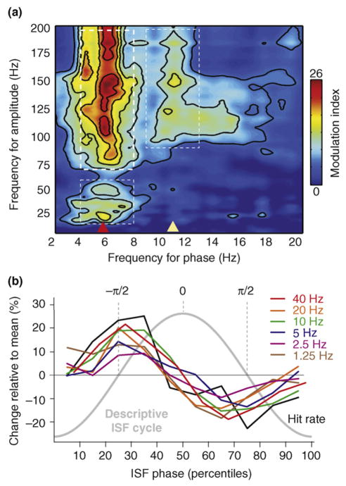
Neuroelectric oscillations reflect rhythmic shifting of neuronal ensembles between high and low excitability states. In natural settings, important stimuli often occur in rhythmic streams, and when oscillations entrain to an input rhythm their high excitability phases coincide with events in the stream, effectively amplifying neuronal input responses. When operating in a 'rhythmic mode', attention can use these differential excitability states as a mechanism of selection by simply enforcing oscillatory entrainment to a task-relevant input stream. When there is no low-frequency rhythm that oscillations can entrain to, attention operates in a 'continuous mode', characterized by extended increase in gamma synchrony. We review the evidence for early sensory selection by oscillatory phase-amplitude modulations, its mechanisms and its perceptual and behavioral consequences.


Figures

Figure 1
Figure 1
(a) (i) Theta-band (5–9 Hz band pass) oscillatory activity from a lower supragranular site in primary auditory cortex (asterisks at left) superimposed on the underlying current source density (CSD) profile for the supregranular layers. Net outward transmembrane current flow generates net extracellular current sources (blue), whereas net inward current flow generates current sinks (red). The theta oscillation at this site represents the ‘underside’ of the superficial current dipole so that negative deflections correspond to current sinks and positive deflections reflect current sources, alternating at a theta rhythm. (ii) Multiunit activity (MUA) simultaneously recorded from the same site. Drop lines are provided to show the relationship between the initial three negative deflections and sinks at this site and MUA correlates. Note that current sinks and sources correspond to MUA peaks and troughs, indicating alternations in local neuronal excitability (adapted from Ref. [18]). (b) Relation between gamma-band (30–90 Hz) oscillatory phase and neuronal firing (MUA) from a recording in macaque visual area V4. Vertical lines at the bottom represent occurrence of action potentials (adapted from Ref. [14]).
Figure 2
Figure 2
Mechanisms of driving and modulatory inputs. (a) Box plots show pooled onset latencies of the characteristic frequency-tone- (aud; blue) and somatosensory-stimulus (som; red)-related CSD response in supragranular (S), granular (G) and infragranular (I) layers across experiments. The boxes have lines at the lower quartile, median and upper quartile values and the notches in boxes graphically show the 95% confidence interval about the median of each distribution. Brackets indicate the significant post hoc comparisons calculated using Games-Howell tests (P < 0.01). (b) Box plots show pooled (n = 38) CSD and MUA amplitudes on the selected channels (S, G and I) averaged for the 15–60 ms time interval for the same conditions as (a), plus the bimodal condition. Brackets indicate the significant post hoc comparisons calculated using Games-Howell tests (P < 0.01). (c) (i) Pooled (n = 38) post-stimulus:pre-stimulus single-trial oscillatory amplitude ratio (0 to 250 ms: −500 to −250 ms) for different frequency intervals (different colors) of the auditory (AU), somatosensory (SS) and bimodal supragranular responses. Stars denote where the amplitude ratio is significantly different across the pre- and post-stimulus periods (one-sample t tests, P < 0.01). (ii) Pooled intertrial coherence (ITC) expressed as a vector quantity (mean resultant length) measured at 15 ms post-stimulus (the time of the initial peak response). Note that in the case of somatosensory events an increase in phase concentration only occurs in the low-delta (1–2.2 Hz), theta (4.8–9.3 Hz) and gamma (25–49 Hz) bands, indicated by colored arrows on the right. (d) Relative distributions and concentrations of calbindin-positive matrix cells (bottom left) and parvalbumin-positive core cells (bottom right) in a frontal section through the middle of a macaque monkey thalamus. The projections of the matrix to superficial layers of cortex over a wide extent and unconstrained by areal borders is shown at the top. Core cells restricted to individual nuclei (e.g. the ventral posterior nucleus) project in a topographically ordered manner to the middle layers of single functional cortical fields. Abbreviations: CL, central lateral nucleus; CM, centre median nucleus; Hl, lateral habenular nuclei; Hm, medial habenular nuclei; LD, lateral dorsal nucleus; LGN, lateral geniculate nucleus; LP, lateral posterior nucleus; MD, mediodorsal nucleus; OT, optic tract; P, color-coded retinal ganglion cells; Pla, anterior pulvinar; PP, peripeduncular nucleus; R, reticular nucleus; s, s laminae; SNr, substantia nigra pars reticularis; VMb, basal ventral medial nucleus; VPi, ventral posterior inferior nucleus; VPM, ventral posterior medial nucleus (figure adapted, with permission, from Ref. [23]).
Figure 3
Figure 3
Effects of task demands on oscillatory dynamics and behavior. (a) Depiction of a vigilance paradigm (i) (adapted, with permission, from Refs [14,29,30]), and a rhythmic stream paradigm (ii) (adapted, with permission, from Ref. [28]). In both cases the subjects make manual responses to target stimuli. The key difference is that stimuli occur randomly in the first case but are arranged in a rhythmic stream in the second. The former suppresses low-frequency oscillatory entrainment, whereas the latter facilitates it. (b) Behavioral correlates of oscillatory modulation by attention in the same two studies. Reaction time (RT) is predicted by gamma-band amplitude in the former (i and ii) and by delta-band phase in the latter (iii and iv). (c) Variations in stimulus discriminability (d′) in a tone discrimination, depending on whether targets occurred in (middle) or out of phase (left and right) with an attended rhythm (adapted, with permission, from Ref. [36]).
Figure 4
Figure 4
Attentional modulation of delta phase and its related cascade of effects. (a,b) Color maps show CSD profiles related to standard visual (Vis) stimuli in the attend visual (AV) and attend auditory (AA) conditions for the −800 to +400 ms time frame from a representative experiment. Red arrow indicates the visual event used as a trigger (0 ms). Blue and red brackets indicate the time frame where adjoining auditory (Aud) and visual events occur; because stimuli are jittered the responses to prior stimuli are somewhat ‘smeared’ over time. (c) Overlay of CSD waveforms from supragranular site (S) in the AV and AA conditions. (d) (i) Time–frequency plot of the average oscillatory amplitude of the wavelet transformed single trials from the supragranular site in (A); note variations in theta (~6 Hz) and gamma (~40 Hz) amplitudes are coupled with stimulus-entrained delta phase. (ii) An overlay of the variations in time course of averaged (37–57 Hz) gamma amplitude in the AV and AA conditions. (e) Pooled (n = 24) normalized gamma amplitude and MUA differences between AV and AA conditions ([AV−AA]/AA) for the −325 to −275 and −50 to 0 ms time frames. Notches in the boxes depict a 95% confidence interval about the median of each distribution.
Figure I
Figure I
(a) Cross-frequency phase-amplitude coupling plot showing that high-gamma-band amplitude (thick-dashed white box) is grouped according to theta-band oscillatory phase (red arrow). More subtle phase-amplitude-coupling effects (thin-dashed white boxes) are also apparent between theta-phase and gamma amplitude, and between ~10 Hz (alpha and mu) band phase (yellow arrow) and high gamma amplitude (adapted, with permission, from Ref. [64]). (b) Based on EEG recorded from a scalp electrode (vertex to mastoid reference), 1–40 Hz oscillation amplitudes are coupled with infraslow frequency (ISF) phase. Amplitude values are represented as a percentage change from the mean for each frequency as a function of ISF phase. ISF phase ranges from −π to +π in 10%-ile bins. Note that the phase difference (mean +/− standard error of the mean) between the amplitude envelope of faster oscillations and ISF is consistently at ~ −π/2. Note also that behavioral responding accuracy (hit rate, black line) is coupled with ISF phase in the same way as the >1 Hz oscillations (adapted, with permission, from Ref. [65]).

References

    1. Bishop G. Cyclical changes in excitability of the optic pathway of the rabbit. Am J Physiol. 1933;103:213–224.
    1. Maurer AP, McNaughten BL. Network and intrinsic cellular mechanisms underlying theta phase precession of hippocampal neurons. Trends Neurosci. 2007;30:325–333. - PubMed
    1. Jensen O, et al. Human gamma-frequency oscillations associated with attention and memory. Trends Neurosci. 2007;30:317–324. - PubMed
    1. Palva S, Palva JM. New vistas for α frequency band oscillations. Trends Neurosci. 2007;30:150–158. - PubMed
    1. Singer W, Gray CM. Visual feature integration and the temporal correlation hypothesis. Annu Rev Neurosci. 1995;18:555–586. - PubMed

Publication types