. 2009 Jan 1;325(1):138-50.
doi: 10.1016/j.ydbio.2008.10.006. Epub 2008 Nov 1.

Neuromancer1 and Neuromancer2 regulate cell fate specification in the developing embryonic CNS of Drosophila melanogaster

Affiliations

Neuromancer1 and Neuromancer2 regulate cell fate specification in the developing embryonic CNS of Drosophila melanogaster

S M Leal et al. Dev Biol. .

Abstract

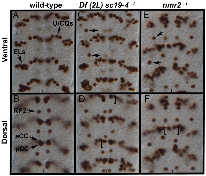
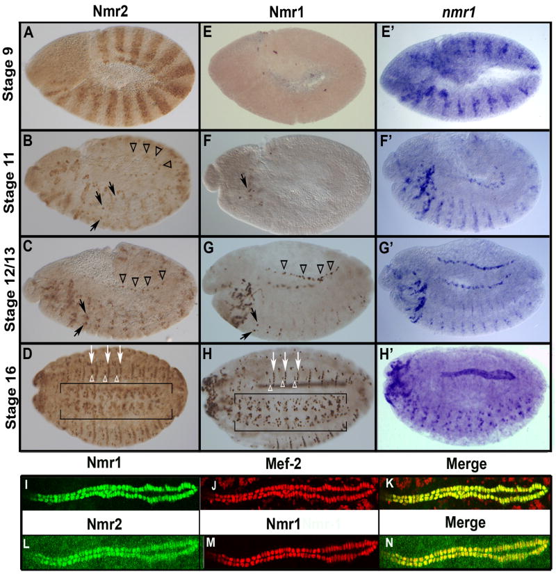
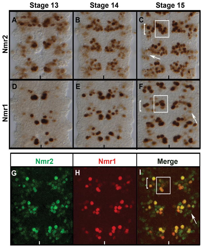
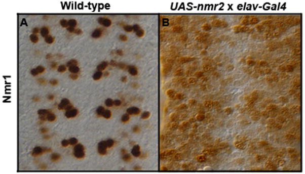
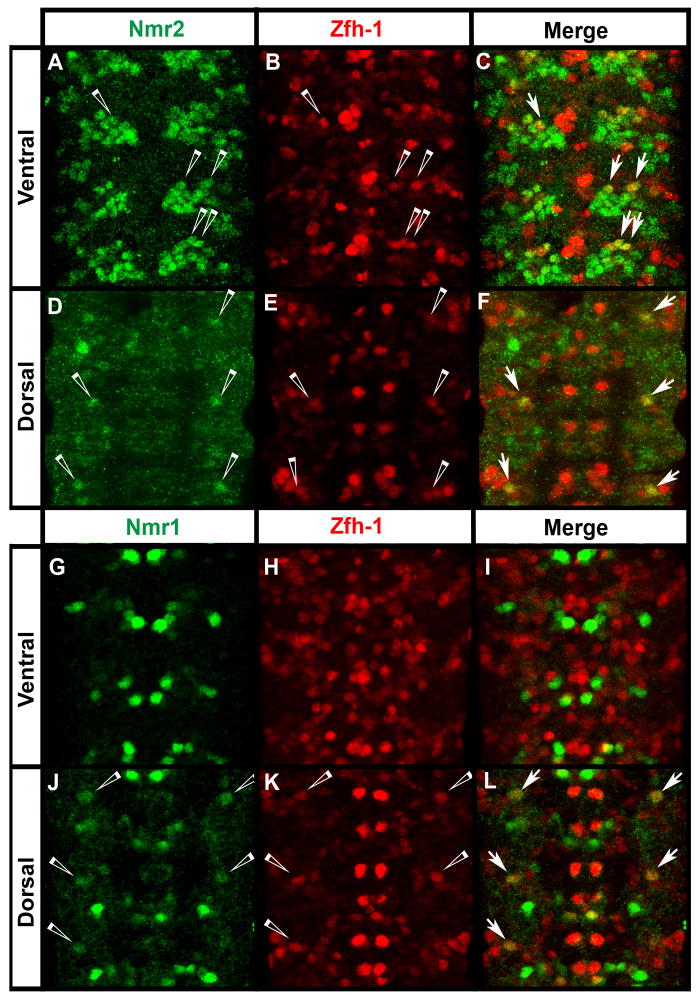
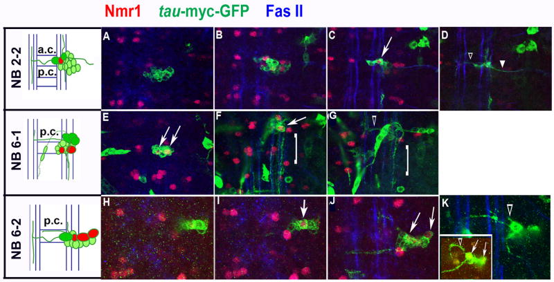
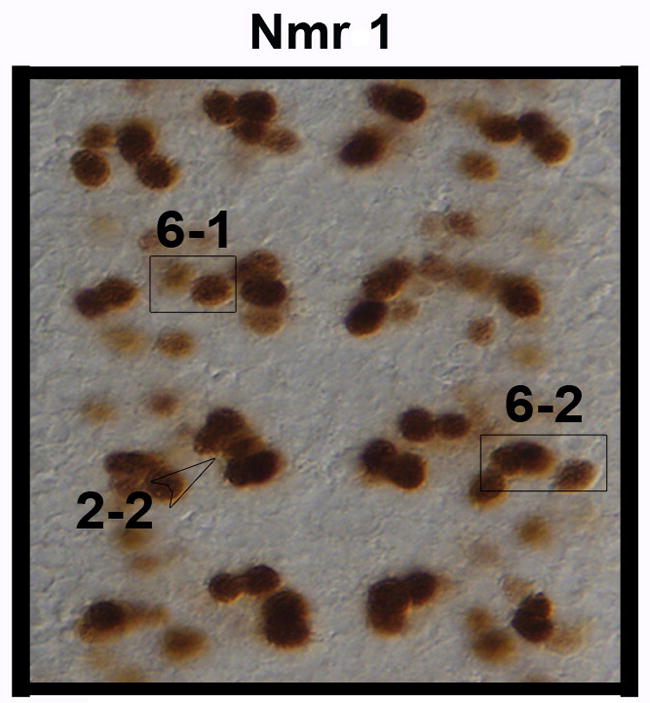
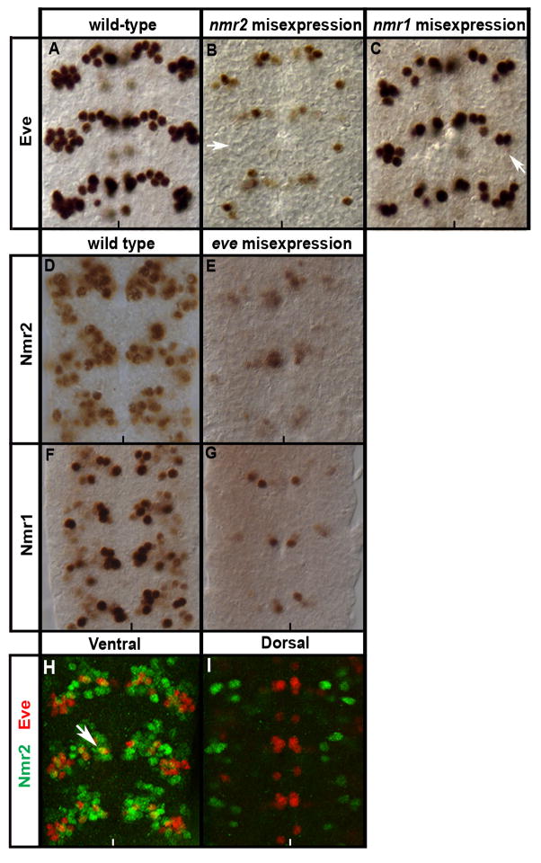
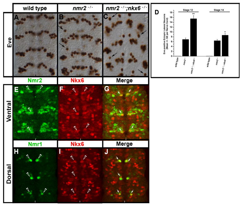
T-box genes encode a large family of transcription factors that regulate many developmental processes in vertebrates and invertebrates. In addition to their roles in regulating embryonic heart and epidermal development in Drosophila, we provide evidence that the T-box transcription factors neuromancer1 (nmr1) and neuromancer2 (nmr2) play key roles in embryonic CNS development. We verify that nmr1 and nmr2 function in a partially redundant manner to regulate neuronal cell fate by inhibiting even-skipped (eve) expression in specific cells in the CNS. Consistent with their redundant function, nmr1 and nmr2 exhibit overlapping yet distinct protein expression profiles within the CNS. Of note, nmr2 transcript and protein are expressed in identical patterns of segment polarity stripes, defined sets of neuroblasts, many ganglion mother cells and discrete populations of neurons. However, while we observe nmr1 transcripts in segment polarity stripes and specific neural precursors in early stages of CNS development, we first detect Nmr1 protein in later stages of CNS development where it is restricted to discrete subsets of Nmr2-positive neurons. Expression studies identify nearly all Nmr1/2 co-expressing neurons as interneurons, while a single Eve-positive U/CQ motor neuron weakly co-expresses Nmr2. Lineage studies map a subset of Nmr1/2-positive neurons to neuroblast lineages 2-2, 6-1, and 6-2 while genetic studies reveal that nmr2 collaborates with nkx6 to regulate eve expression in the CNS. Thus, nmr1 and nmr2 appear to act together as members of the combinatorial code of transcription factors that govern neuronal subtype identity in the CNS.


Figures

Figure 1
Figure 1
nmr2 represses eve expression in the Drosophila nerve cord. Stage 15 wild-type (A,B), Df(2L)sc19-4−/− (C,D), and nmr2−/− (E,F) mutant nerve cords stained for Eve. (A,B) In wild-type nerve cords Eve labels 5–6 U/CQ, 10 EL, RP2, and aCC/pCC neurons as indicated. (C) In Df(2L)sc19-4−/− mutant embryos, ectopic Eve-positive neurons arise laterally (arrows) and (D) there is a decrease in the presence of Eve-positive aCC/pCC neurons (brackets). (E,F) nmr2−/− mutant embryos (midGA174) exhibit the same phenotype as that observed in Df(2L)sc19-4−/− embryos. Panels A, C, E – ventral views and panels B, D, F – dorsal views of the Drosophila nerve cord; the vertical line marks the midline.
Figure 2
Figure 2
Nmr1 and Nmr2 protein expression during embryogenesis. Nmr2 (A–D,L), Nmr1 (E–H,I) and nmr1 mRNA transcript (E′–H′) expression in wild-type embryos at progressively later stages of development (A) Stage 9 embryos express Nmr2 in segment polarity stripes. (B) During early stage 12, Nmr2 is expressed in subsets of neuroblasts and their progeny in the CNS (arrows) as well as in heart precursor cells in the dorsal mesoderm (arrowheads). (C) During late stage 12, Nmr2 is expressed prominently in a medial pair of CNS neurons in each segment (arrows) and is also expressed in heart precursor cells (arrowheads). (D) By stage 16 Nmr2 is expressed in many neurons in the CNS (brackets), narrower transverse stripes of ectodermal cells (white arrows), and clusters of ectodermal cells (white arrowheads) between these stripes. (E) Stage 9 embryos do not express Nmr1 protein while (E′) nmr1 transcripts are clearly detected in segment polarity stripes. (F) Nmr1 protein is first detected during early stage 12 in a few cells in the cephalic region (arrow), (F′) while similarly staged embryos express nmr1 transcripts in segment polarity stripes, neuronal precursors and neurons. (G, G′) By stage 13, Nmr1 protein and RNA are expressed in identical patterns of CNS neurons (arrows) and heart precursors in the mesoderm (arrowheads). (H, H′) By stage 16, Nmr1 protein and RNA are co-expressed in many neurons in the CNS (H, brackets), all cardioblasts as well as in narrow stripes of ectodermal cells (H, white arrows). (I–N) High magnification views of the Drosophila heart at stage 16 show that Nmr1 (I, green; M, red) and Nmr2 (L, green) are co-expressed with Mef-2 (J, red) in all cardioblasts (K,N). Whole mounts in the lateral orientation; Anterior, left; Posterior, right; Dorsal, up; Ventral, down.
Figure 3
Figure 3
Co-expression of Nmr2 and Nmr1 in the Drosophila CNS. Stage 13 (A,D,) stage 14 (B,E), and late stage 15 (C,F,G–I) wild-type embryos labeled for Nmr2 (A–C, G, I), Nmr1 (D–F, H, I) and co-labeled for Nmr1 and Nmr2 (I). (A) During stage 13 Nmr2 is expressed strongly in a cluster of medial neurons and weakly in a group of lateral neurons. (B) By stage 14, Nmr2 is expressed strongly in a cluster of approximately 10–15 cells per hemisegment with enhanced expression in the medial and intermediate regions of the nerve cord and diminished expression laterally. (C) This pattern of expression is prominently defined in stage 15 where Nmr2 is expressed strongly in a cluster of approximately 8 medial neurons (box) and 8 lateral neurons (bracket). Nmr2 is also expressed weakly in about 5–6 neurons within the lateral cluster (arrow). (D) During stage 13 Nmr1 is expressed strongly in a small number of medial neurons and expressed weakly in a few neurons in the intermediate region. (E) By stage 14, Nmr1 expression remains high in medial neurons and expression is now observed in additional neurons in the intermediate regions of the CNS. (F) By stage 15, Nmr1 is expressed in many discrete sets of neurons in the CNS: a medial cluster of approximately 3–5 neurons (box), a lateral cluster of 7–8 neurons (bracket), and a few dispersed lateral neurons (arrow). (G–I) Double-labeling for Nmr2 (green) and Nmr1 (red) in stage 15 embryos indicates that a subset of Nmr2-positive neurons express Nmr1 (merge).
Figure 4
Figure 4
Nmr2 enhances Nmr1 expression. Stage 15 embryonic nerve cords stained for Nmr1. (A) Wild-type expression pattern of Nmr1 in the ventral nerve cord. (B) Misexpression of Nmr2 in all post-mitotic neurons (ElavGAL4::UAS-nmr2) significantly enhances Nmr1 expression throughout the nerve cord.
Figure 5
Figure 5
Nmr1 and Nmr2 are detected in a few Zfh-1-expressing motor neurons. (A–L) Wild-type stage 15 embryos stained for Zfh-1 (red) and Nmr2 (green) or Nmr1 (green). (A–C) In the ventral region of the CNS, Nmr2-positive and Zfh-1-positive neurons are co-expressed in approximately 2–3 neurons in the medial Nmr2-expressing cluster per hemisegment (merge, arrows). (D–F) Dorsally in the CNS, a single lateral neuron co-expresses Nmr2 and Zfh-1 per hemisegment (merge, arrows). (G–I) In the ventral region of the CNS, no cells co-express Nmr1 (green) and Zfh-1 (red). (J–L) In the dorsal region of the CNS, one lateral neuron weakly co-expresses Nmr1 and Zfh-1 per hemisegment (merge, arrows).
Figure 6
Figure 6
A subset of Nmr1/−2 expressing interneurons derived from neuroblast lineages 2-2, 6-1, and 6-2. Cartoons detail the positions and axon morphologies of Nmr1-positive interneurons (red) within identified NB clones in the CNS of wild-type, stage 15–17 embryos. The most ventral neurons are shaded dark green. Neurons are drawn in relation to the anterior and posterior commissures (AC/PC) and the longitudinal connectives (blue). From top (dorsal) to bottom (ventral) the compressed confocal images represent ~1 μM thick sections of Nmr1-positive clones. The nerve cord is triple-labeled for Nmr1 (red), axons (green, anti-GFP), and longitudinal connectives (blue, anti-FasII). (A–D) A lineage clone that contains one Nmr1-positive neuron (arrow) extends axons contralaterally (D, open arrowhead) and a motor neuron axon branch (D, closed arrowhead). (E–G) A lineage clone that contains two Nmr1-positive interneurons (white arrows) extends a collateral axon branch (G, open arrowheads) and a cascade of ipsilateral axons (F,G, brackets). (H–K) A lineage clone that contains three Nmr1-positive neurons (arrows) extends a small ipsilateral axon branch (J, open arrowhead) as well as anterior and posterior collateral axons. Occasionally, NB 6-2 clones exhibit an ipsilateral axon branch with a prominent arch and this axonal phenotype was detected in a similar clone (K, inset).
Figure 7
Figure 7
Expression pattern of Nmr1-positive neurons derived from NBs 2-2, 6-1 and and 6-2. The boxed sets of neurons highlight the Nmr1-positive neurons derived from NB lineages 6-1 (left) and 6-2 (right). An open arrowhead points to the Nmr1-positive neuron derived from the NB 2-2 lineage.
Figure 8
Figure 8
A subset of medial Nmr2 and Nmr1 interneurons co-express glutamic acid decarboxylase. (A–C) Stage 15 nerve cords from the H15-LacZ reporter line, which labels all Nmr1-positive and Nmr2-positive neurons. (A) Staining for glutamic acid decarboxylase (Gad) specifically identifies GABAergic populations of interneurons (green). (B) Staining for β-Galactosidase (B-Gal) marks all Nmr1 and Nmr2 positive neurons (red). (C) A prominent, medial pair of previously identified Nmr1 and Nmr2 co-expressing neurons detected within each segment of the nerve cord accumulates a high level of vesicular Gad at the plasma membrane (arrows, merge).
Figure 9
Figure 9
Nmr1 and Nmr2 repress Eve expression. Wild-type stage 15 embryonic nerve cords stained for Eve (A–C, H,I), Nmr2 (D,E,H,I), and Nmr1 (F,G). (A) Wild-type expression pattern of Eve within the ventral region of the CNS. (B) Misexpression of Nmr2 in all post-mitotic neurons (ElavGAL4::UAS-nmr2) significantly reduces Eve expression in all Eve-positive neurons (arrow points to location of ELs). (C) Misexpression of Nmr1 throughout the CNS (ElavGAL4::UAS-nmr1) causes a mild decrease of Eve expression (arrow points to ELs). (D,F) Wild-type expression pattern of Nmr2 (D) and Nmr1 (F). (E,G) Expression of Eve throughout the CNS (ElavGAL4::UAS-eve) results in a significant decrease of Nmr2 (E) and Nmr1 (G). (H,I) Eve (red) exhibits a predominantly mutually exclusive expression pattern with Nmr2 (green) in the ventral (H) and dorsal (I) regions of the nerve cord while a single Eve-positive U/CQ neuron (H) co-expresses Nmr2 (arrows).
Figure 10
Figure 10
nmr2 and nkx6 collaborate to inhibit eve expression. (A–C) Stage 14 wild-type and mutant embryonic nerve cords stained for Eve, (D) a chart quantifies the number of Eve-positive lateral neurons in wild-type, nmr2−/−, and nmr2−/−;nkx6−/− mutant backgrounds at stages 12 and 14, (E–J) immunofluorescent staining of Nmr2-, Nmr1-, and Nkx-6-positive neurons in the CNS of Stage 15 wild-type embryos. All images depict the ventral region of the CNS. (A) Stage 14 wild-type Eve expression pattern. (B) As shown previously, nmr2−/− mutant embryos exhibit Eve-positive ectopic lateral neurons (arrows, see Fig. 1B). (C) nmr2−/−;nkx6−/− double mutant embryos exhibit increased numbers of Eve-positive ectopic lateral neurons throughout the nerve cord as compared with nmr2−/− mutants (arrows). The nerve cords of nmr2−/−;nkx6−/− mutants also exhibit severe orientation defects that become pronounced at stages 15–16 (data not shown). (D) Each bar value represents the mean of 20 embryos scored for the number of Eve-positive ectopic lateral neurons observed throughout the nerve cord at stages 12 and 14. The error bars represent the S.E.M. and statistical significance was determined by One-way Analysis of Variance (ANOVA) where p** < 0.001 and p* < 0.01. (E–G) Nmr2 (green) and Nkx6 (red) exhibit a mutually exclusive expression pattern with the exception of one medial neuron per hemisegment that co-expresses Nmr2 and Nkx6 (merge, arrows). (H–J) Nmr1 (green) and Nkx6 (red) are also co-expressed in one medial neuron per hemisegment.

Similar articles

Cited by

References

    1. Adell T, Muller WE. Expression pattern of the Brachyury and Tbx2 homologues from the sponge Suberites domuncula. Biol Cell. 2005;97:641–650. - PubMed
    1. Ahn DG, Ruvinsky I, Oates AC, et al. tbx20, a new vertebrate T-box gene expressed in the cranial motor neurons and developing cardiovascular structures in zebrafish. Mech Dev. 2000;95:253–258. - PubMed
    1. Alexandre C, Lecourtois M, Vincent J. Wingless and Hedgehog pattern Drosophila denticle belts by regulating the production of short-range signals. Development. 1999;126:689–98. - PubMed
    1. Basson CT, Huang T, Lin RC, et al. Different TBX5 interactions in heart and limb defined by Holt-Oram syndrome mutations. Proc Natl Acad Sci USA. 1999;96:2919–2924. - PMC - PubMed
    1. Bossing T, Brand AH. Determination of cell fate along the anteroposterior axis of the Drosophila ventral midline. Development. 2006;133:1001–1012. - PubMed

Publication types

MeSH terms