. 2008 Nov 14;135(4):679-90.
doi: 10.1016/j.cell.2008.09.038.

Mistranslation of membrane proteins and two-component system activation trigger antibiotic-mediated cell death

Affiliations

Mistranslation of membrane proteins and two-component system activation trigger antibiotic-mediated cell death

Michael A Kohanski et al. Cell. .

Abstract

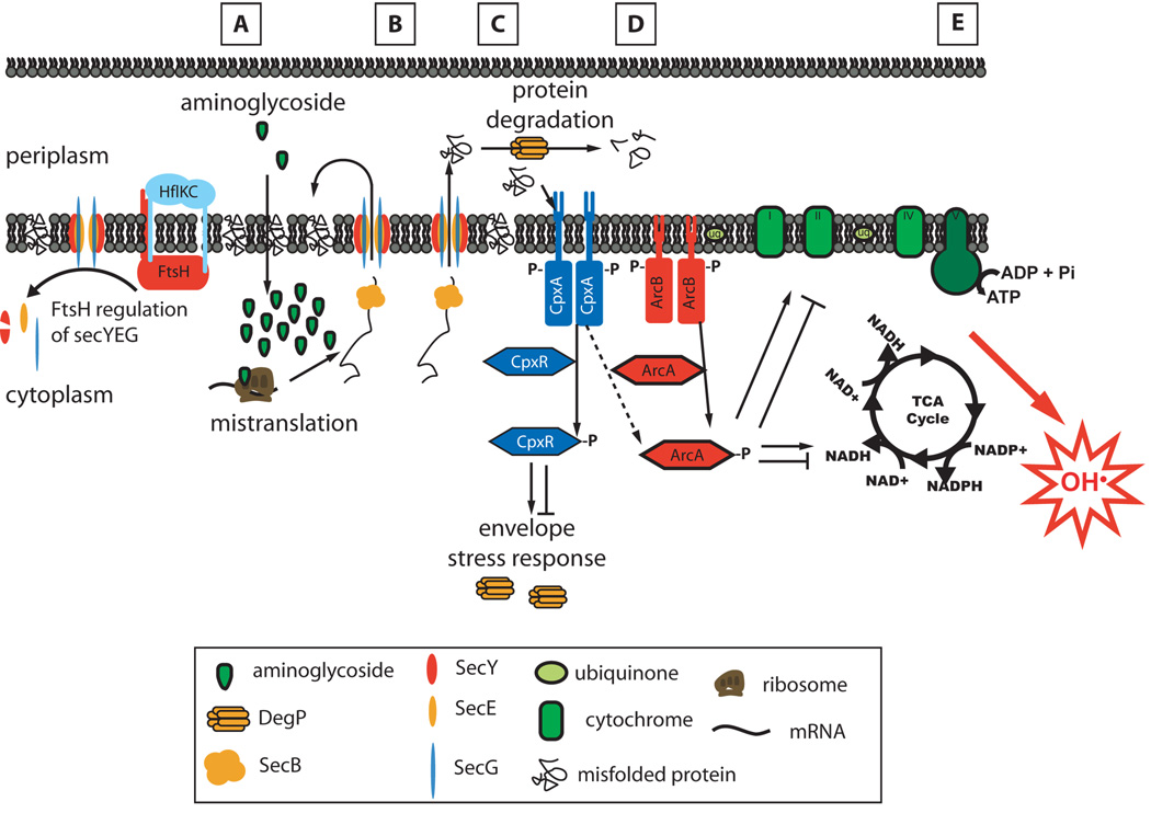
Aminoglycoside antibiotics, such as gentamicin and kanamycin, directly target the ribosome, yet the mechanisms by which these bactericidal drugs induce cell death are not fully understood. Recently, oxidative stress has been implicated as one of the mechanisms whereby bactericidal antibiotics kill bacteria. Here, we use systems-level approaches and phenotypic analyses to provide insight into the pathway whereby aminoglycosides ultimately trigger hydroxyl radical formation. We show, by disabling systems that facilitate membrane protein traffic, that mistranslation and misfolding of membrane proteins are central to aminoglycoside-induced oxidative stress and cell death. Signaling through the envelope stress-response two-component system is found to be a key player in this process, and the redox-responsive two-component system is shown to have an associated role. Additionally, we show that these two-component systems play a general role in bactericidal antibiotic-mediated oxidative stress and cell death, expanding our understanding of the common mechanism of killing induced by bactericidal antibiotics.


Figures

Figure 1
Figure 1
Identification of pathways related to aminoglycoside lethality. Significantly changing genes from a comparison of treatment with a lethal aminoglycoside versus the bacteriostatic ribosome inhibitor, spectinomycin, were filtered through an E. coli gene connectivity map generated using the Context Likelihood of Relatedness algorithm (Faith et al., 2007); the resultant gene networks were analyzed for functional enrichment. The strains exhibiting decreased growth or increased growth found from a high-throughput screen of an E. coli single-gene deletion library treated with gentamicin were overlaid on these networks.
Figure 2
Figure 2
CLR-based networks of genes whose expression changed significantly due to aminoglycoside (gentamicin or kanamycin) treatment. In this figure, upregulated gene expression is shown in green, downregulated gene expression is in yellow, and non-perturbed gene expression is shown in light blue. Overlaid on these networks are the outliers from a screen of the single-deletion knockout (KO) library grown in the presence of 5µg/ml gentamicin (increased growth of KO, red rim; decreased growth of KO, blue rim; no change in growth of KO, black rim). We note that no single-knockouts of genes in these networks show decreased growth in the presence of gentamicin. (A) Network of genes showing pathway enrichment for the electron transport chain (ETC), tricarboxylic acid (TCA) cycle and respiration (p < 10−11), and transcription factor enrichment for metabolic regulators including ArcA (p = 1.8×10⁻¹²). (B) Network of genes showing pathway enrichment for the response to misfolded proteins (p = 1×10⁻¹⁷) and the heat shock sigma factor, σH (RpoH: p = 1.2×10⁻¹¹).
Figure 3
Figure 3
Disrupting membrane regulatory systems enhances aminoglycoside lethality. Percent survival of wildtype E. coli (black squares), ΔsecG (red circles), ΔhflK (green triangles) and ΔhflC (blue triangles), following treatment with (A) 5µg/ml gentamicin, (B) 100ng/ml norfloxacin, or (C) 3µg/ml ampicillin. Mean +/− S.E.M. are shown for all figures.
Figure 4
Figure 4
Disrupting membrane regulatory systems and the envelope stress response enhances aminoglycoside-induced hydroxyl radical formation and membrane depolarization. (A–D) Fluorescence for each strain relative to the maximum fluorescence achieved in the wildtype background using (A,C) the hydroxyl radical detecting dye, HPF, or (B,D) the membrane depolarization dye, DIBAC4(3). (A) Hydroxyl radical formation and (B) membrane depolarization of wildtype E. coli (black squares), ΔsecG (red circles), ΔhflK (green triangles) and ΔhflC (blue triangles), following treatment with 5µg/ml gentamicin. (C) Hydroxyl radical formation and (D) membrane depolarization of wildtype E. coli (black squares), ΔdegP (red diamonds), ΔcpxA (green diamonds), ΔcpxR (red diamonds), and ΔarcA (blue circles), following treatment with 5µg/ml gentamicin. (E) Fold-change gene expression relative to baseline (t = 0min) of the envelope stress response genes, cpxP (sqaures) and degP (triangles), for wildtype E. coli (black), ΔcpxR (brown) and DcpxA(green) following treatment with 5µg/ml gentamicin.
Figure 5
Figure 5
The envelope stress response and redox-responsive two-component systems are involved in bactericidal drug-mediated lethality. Percent survival of wildtype E. coli (black squares), ΔdegP (red diamonds), ΔcpxA (green diamonds), ΔcpxR (red diamonds), and ΔarcA (blue circles), following treatment with (A) 5µg/ml gentamicin, (B) 100ng/ml norfloxacin, or (C) 3mg/ml ampicillin.
Figure 6
Figure 6
Envelope stress response and redox-responsive two-component systems trigger hydroxyl radical formation due to mistranslation of membrane proteins. Survival, hydroxyl radical formation and membrane depolarization of wildtype E. coli (black squares), ΔhflKΔcpxA (blue triangles), ΔhflKΔarcA (brown triangles), ΔsecGΔcpxA (red circles) and ΔsecGΔarcA (green circles), following treatment with 5µg/ml gentamicin. (A) Percent survival of each strain relative to when gentamicin treatment started (0 hours). (B and C) Fluorescence for each strain relative to the maximum fluorescence achieved in the wildtype background using (B) the hydroxyl radical detecting dye, HPF, or (C) the membrane depolarization dye, DIBAC4(3).
Figure 7
Figure 7
Proposed mechanism by which aminoglycosides trigger hydroxyl radical formation and cell death. (A) The primary interaction between the aminoglycoside and ribosome causes protein mistranslation. (B) Mistranslated, immature membrane proteins are brought to membrane translocation complexes (e.g., SecYEG) by chaperones proteins (SecB), and are translocated across the inner membrane into the periplasmic space or inserted into the membrane. (C) Due to mistranslation, many of these proteins are misfolded, leading to phosphorylation of CpxA. (D) Activated CpxA phosphorylates CpxR, which upregulates expression of envelope stress response proteins, such as the periplasmic protease DegP. CpxA may also activate ArcA, which regulates a large number of metabolic and respiratory genes. These changes shift the cell into a state that (E) provokes free radical formation, ultimately culminating in hydroxyl radical formation and cell death. We found that β-lactams and quinolones also trigger hydroxyl radical formation and cell death through the Cpx and Arc two-component systems (D, E); the specific triggers for β-lactams and quinolones remain to be worked out.

References

    1. Akiyama Y, Kihara A, Tokuda H, Ito K. FtsH (HflB) is an ATP-dependent protease selectively acting on SecY and some other membrane proteins. J Biol Chem. 1996;271:31196–31201. - PubMed
    1. Ashburner M, Ball CA, Blake JA, Botstein D, Butler H, Cherry JM, Davis AP, Dolinski K, Dwight SS, Eppig JT, et al. Gene ontology: tool for the unification of biology. The Gene Ontology Consortium. Nat Genet. 2000;25:25–29. - PMC - PubMed
    1. Baba T, Ara T, Hasegawa M, Takai Y, Okumura Y, Baba M, Datsenko KA, Tomita M, Wanner BL, Mori H. Construction of Escherichia coli K-12 in-frame, single-gene knockout mutants: the Keio collection. Mol Syst Biol. 2006;2 2006.0008. - PMC - PubMed
    1. Bolstad BM, Irizarry RA, Astrand M, Speed TP. A comparison of normalization methods for high density oligonucleotide array data based on variance and bias. Bioinformatics. 2003;19:185–193. - PubMed
    1. Boyle EI, Weng S, Gollub J, Jin H, Botstein D, Cherry JM, Sherlock G. GO::TermFinder--open source software for accessing Gene Ontology information and finding significantly enriched Gene Ontology terms associated with a list of genes. Bioinformatics. 2004;20:3710–3715. - PMC - PubMed

Publication types

LinkOut - more resources