. 2009 Feb;215(2):243-52.
doi: 10.1016/j.expneurol.2008.10.005. Epub 2008 Oct 26.

Posttraumatic epilepsy after controlled cortical impact injury in mice

Affiliations

Posttraumatic epilepsy after controlled cortical impact injury in mice

Robert F Hunt et al. Exp Neurol. 2009 Feb.

Abstract

Many patients develop temporal lobe epilepsy after trauma, but basic mechanisms underlying the development of chronic seizures after head injury remain poorly understood. Using the controlled cortical impact injury model we examined whether mice developed spontaneous seizures after mild (0.5 mm injury depth) or severe (1.0 mm injury depth) brain injury and how subsequent posttraumatic mossy fiber sprouting was associated with excitability in the dentate gyrus 42-71 d after injury. After several weeks, spontaneous behavioral seizures were observed in 20% of mice with mild and 36% of mice with severe injury. Mossy fiber sprouting was typically present in septal slices of the dentate gyrus ipsilateral to the injury, but not in control mice. In slices with mossy fiber sprouting, perforant path stimulation revealed a significant reduction (P<0.01) in paired-pulse ratios in dentate granule cells at 20 ms and 40 ms interpulse intervals, but not at 80 ms or 160 ms intervals. These slices were also characterized by spontaneous and hilar-evoked epileptiform activity in the dentate gyrus in the presence of Mg(2+)-free ACSF containing 100 microM picrotoxin. In contrast, paired-pulse and hilar-evoked responses in slices from injured animals that did not display mossy fiber sprouting were not different from controls. These data suggest the development of spontaneous posttraumatic seizures as well as structural and functional network changes associated with temporal lobe epilepsy in the mouse dentate gyrus by 71 d after CCI injury. Identifying experimental injury models that exhibit similar pathology to injury-induced epilepsy in humans should help to elucidate the mechanisms by which the injured brain becomes epileptic.


Figures

Figure 1
Figure 1
Reorganization of mossy fiber projections after severe injury. (A) Representative Timm and Nissl counter-stained coronal section (~Bregma −2.0mm) from a mouse after severe controlled cortical impact TBI. (B) Timm and Nissl stained horizontal section from a control mouse. (C) Timm and nissl stained section from the contralateral hemisphere of an injured mouse 42d post CCI. (D) Staining in the septal dentate gyrus ipsilateral to the lesion revealed mossy fiber sprouting into the inner molecular layer (arrows) at the same septotemporal level and in the same mouse as in C. (E–G) Higher power images of B–D. Scale bars: 500μm in A, 100μm in B–D, 25μm E–G.
Figure 2
Figure 2
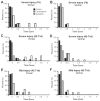
Mossy fiber sprouting progresses with time after CCI injury. Sprouting was typically confined to the septal dentate gyrus of the ipsilateral hemisphere. Timm score ranges are given for three groups (controls, injured ipsilateral hemisphere, and injured contralateral hemisphere) from septal (A) and temporal (B) dentate gyrus 7d after severe injury, septal (C) and temporal (D) dentate gyrus 42–71d after severe injury, and septal (D) and temporal (E) dentate gyrus 42–71d after mild injury.
Figure 3
Figure 3
Paired-pulse ratios in the dentate gyrus are reduced in slices from injured mice with mossy fiber sprouting 42–71d after injury. (A) Representative field-potential recordings evoked by paired-pulsed stimulation of perforant path input to the dentate gyrus at 20, 40, 80, and 160ms interpulse intervals in controls, CCI injured mice with no mossy fiber sprouting (MFS), and CCI injured mice with MFS. Facilitation of the second population spike was observed at all intervals in controls and injured mice without mossy fiber sprouting. Paired-pulse ratios were significantly decreased in slices from injured mice with mossy fiber sprouting at early (20ms and 40ms) but not later (80ms and 160ms) intervals. (B) Bar graph showing the mean PPR for all slices at 20, 40, 80, and 160 ms (n=15–46). Bars represent means ± S.E.M. Asterisks (*) represent a significant reduction in paired-pulse ratios as determined by one-way ANOVA with Tukey’s post hoc analysis (P < 0.01).
Figure 4
Figure 4
Electrical stimulation of mossy fibers in the hilus evokes increased population responses in the dentate gyrus of mice with mossy fiber reorganization 42–71d after CCI injury. (A, B) Hilar-evoked responses in controls and injured mice without mossy fiber sprouting consisted of a single population spike in normal ACSF and in Mg2+-free ACSF containing PTX. (C) Evoked responses in a slice from a CCI-injured mouse with moderate mossy fiber sprouting. A single population spike was evoked in normal ACSF. Multiple population spikes were evoked in Mg2+-free ACSF containing PTX. (D) Hilar stimulation evoked a single population spike in normal ACSF in a slice with robust mossy fiber sprouting. In Mg2+-free ACSF containing PTX, a prolonged negative field-potential shift with secondary population activity was evoked. (D) The underlined portion of the trace in D is expanded below to demonstrate the initial population spike followed by secondary activity. Insets provide representative Timm’s stains demonstrating the level of mossy fiber sprouting typical of slices that produced each response.
Figure 5
Figure 5
Increased excitability is revealed in the dentate gyrus of CCI-injured mice during disinhibition. (A) Hilar stimulation in normal ACSF evoked a single population spike in nearly all slices from injured and control animals. (B) Average Timm scores for slices in which a single population spike, multiple population spikes, or prolonged negative field shifts were evoked in the presence of Mg2+-free ACSF containing PTX. Bars represent means ± S.E.M. Single asterisks (*) represent P < 0.01 and double asterisks (**) represent a P < 0.001 as determined by Kruskal-Wallis with Dunn’s post hoc analysis. (C) Relative range of hilar-evoked population responses in Mg2+-free ACSF containing PTX.
Figure 6
Figure 6
Spontaneous population activity during disinhibition in slices from control and injured mice. (A) Field-potential recordings in the dentate gyrus of a control mouse demonstrate spontaneous positive-going field potential deflections. (B) Spontaneous epileptiform activity consisting of large amplitude negative going field shifts and spontaneous population spikes observed in the dentate gyrus of a mouse with mossy fiber sprouting. Arrows in A and B indicate expanded portions of the respective traces.

Similar articles

Cited by

References

    1. Anderson KJ, Miller KM, Fugaccia I, Scheff SW. Regional distribution of Fluoro-Jade B staining in the hippocampus following traumatic brain injury. Exp Neurol. 2005;193:125–130. - PubMed
    1. Anderson RW, Brown CJ, Blumbers PC, McLean AJ, Jones NR. Impact mechanics and axonal injury in a sheep model. J Neurotrauma. 2003;20:961–974. - PubMed
    1. Annegers JF, Hauser A, Coan SP, Rocca WP. A population-based study of seizures and traumatic brain injuries. N Eng J Med. 1998;338:20–24. - PubMed
    1. Ben-Ari Y. Limbic seizure and brain damage produced by kainic acid: mechanisms and relevance to human temporal lobe epilepsy. Neurosci. 1985;14:375–403. - PubMed
    1. Buckmaster PS, Zhang GF, Yamawaki R. Axon sprouting in a model of temporal lobe epilepsy creates a predominantly excitatory feedback circuit. J Neurosci. 2002;22:6650–8. - PMC - PubMed

Publication types