. 2009 Jan 20;383(2):237-47.
doi: 10.1016/j.virol.2008.10.029. Epub 2008 Nov 14.

Requirements for cell rounding and surface protein down-regulation by Ebola virus glycoprotein

Affiliations

Requirements for cell rounding and surface protein down-regulation by Ebola virus glycoprotein

Joseph R Francica et al. Virology. .

Abstract

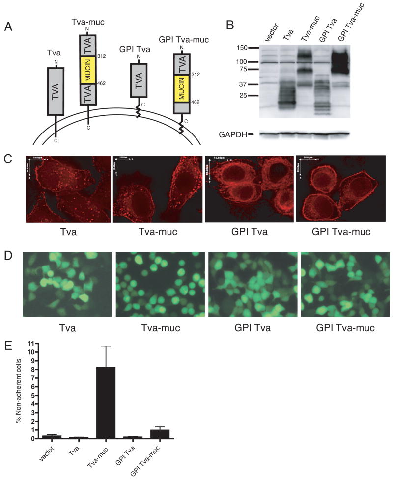
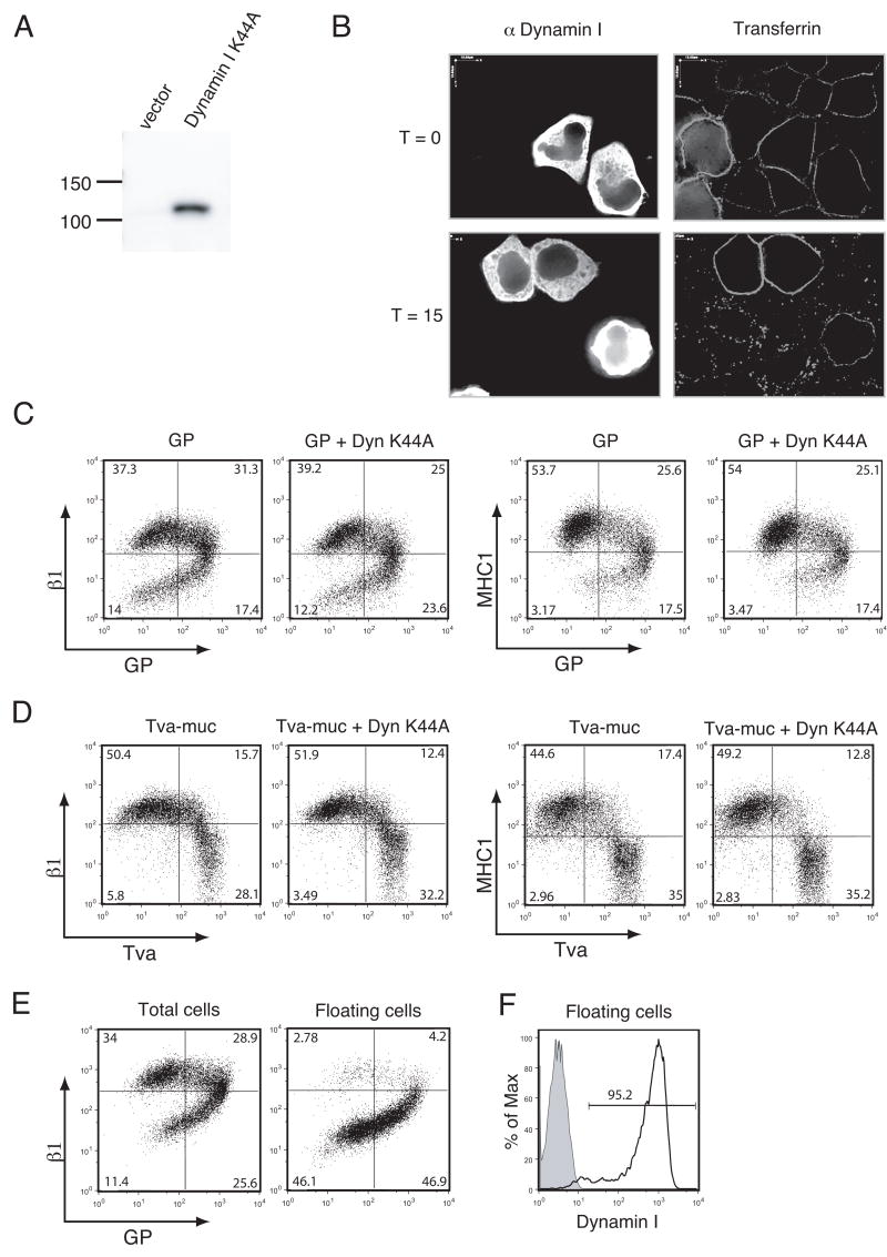
Ebola virus causes an acute hemorrhagic fever that is associated with high morbidity and mortality. The viral glycoprotein is thought to contribute to pathogenesis, though precise mechanisms are unknown. Cellular pathogenesis can be modeled in vitro by expression of the Ebola viral glycoprotein (GP) in cells, which causes dramatic morphological changes, including cell rounding and surface protein down-regulation. These effects are known to be dependent on the presence of a highly glycosylated region of the glycoprotein, the mucin domain. Here we show that the mucin domain from the highly pathogenic Zaire subtype of Ebola virus is sufficient to cause characteristic cytopathology when expressed in the context of a foreign glycoprotein. Similarly to full length Ebola GP, expression of the mucin domain causes rounding, detachment from the extracellular matrix, and the down-regulation of cell surface levels of beta1 integrin and major histocompatibility complex class 1. These effects were not seen when the mucin domain was expressed in the context of a glycophosphatidylinositol-anchored isoform of the foreign glycoprotein. In contrast to earlier analysis of full length Ebola glycoproteins, chimeras carrying the mucin domains from the Zaire and Reston strains appear to cause similar levels of down-modulation and cell detachment. Cytopathology associated with Ebola glycoprotein expression does not occur when GP expression is restricted to the endoplasmic reticulum. In contrast to a previously published report, our results demonstrate that GP-induced surface protein down-regulation is not mediated through a dynamin-dependent pathway. Overall, these results support a model in which the mucin domain of Ebola GP acts at the cell surface to induce protein down modulation and cytopathic effects.


Figures

FIG. 1
FIG. 1
Ebola virus GP-mucin domain is sufficient to induce cell rounding and detachment. (A) Diagram of Tva constructs used to express Ebola Zaire GP-mucin domain. Numbers indicate amino acid position starting from the initial methionine. (B) 239T cells were transfected with pCAGGS alone (vector) or pCAGGS encoding the Tva constructs described in (A). Lysates were harvested in RIPA buffer after 24 h and subjected to SDS-4 to 15% PAGE, transferred to PVDF, and immunoblotted with polyclonal rabbit anti-Tva sera or GAPDH-specific monoclonal antibodies on blots run in parallel. (C) HeLa cells were transfected with Tva constructs. 48 h posttransfection, cells were fixed, permeabilized, and stained for Tva with polyclonal rabbit anti-Tva sera followed by Alexa 594 conjugated antibodies. Z-sections were captured on a fluorescence microscope and deconvoluted with software. Images shown are single, deconvoluted Z-sections. Scale bars are 10.6 μm. (D) 293T cells were co-transfected with Tva constructs and a vector encoding eGFP in a 3:1 ratio. After 24 h fluorescent images were captured on an inverted microscope using a GFP filter. Fields represent findings from multiple experiments. (E) 239T cells were transfected with pCB6 vector alone or pCB6 encoding the Tva constructs described in (A) and co-transfected with a vector encoding eGFP in a 3:1 ratio. 24 h post-transfection, adherent and non-adherent cells were removed. CFP positive cells were counted; data is shown as % non-adherent cells. Graph shows mean of 3 replicates; error bars indicate SD. Results are representative of 2 independent experiments.
FIG. 2
FIG. 2
Surface protein down-regulation by Ebola GP-mucin domain. 239T cells were transfected with vector alone or vector encoding the Tva constructs described in Fig. 1A. Floating and adherent cells were harvested 24 h after transfection, pooled, and stained with polyclonal rabbit anti-Tva sera and FITC-labeled secondary antibodies, co-stained for β1 integrin or MHC1 with PE-Cy5 conjugated monoclonal antibodies and assayed by flow cytometry. Analysis is shown for events in the live cell gate. (A) β1 integrin vs. Tva surface expression. (B) MHC1 vs. Tva surface expression. (C) Histogram representation of β1 integrin surface expression (left panels) and MHC1 surface expression (right panels). Control samples are shown shaded; Tva or GPI Tva is shown as a dashed line; Tva-muc or GPI Tva-muc is shown as a solid line. Data is representative of multiple independent experiments.
FIG. 3
FIG. 3
Ebola virus GP does not round cells when restricted to the ER. (A) 293T cells were transfected with vector alone, vector encoding for GP, or GP-kk (ER-restricted). Lysates were harvested in RIPA buffer after 24 h and subjected to SDS-4 to 15% PAGE, transferred to PVDF, and immunoblotted with polyclonal rabbit anti-GP sera and GAPDH-specific monoclonal antibodies. (B) GP and GP-kk lysates were denatured and incubated with enzyme buffer alone or buffer with PNGase F or Endoglycosidase Hf (at normal or 3x concentration), then subjected to SDS-PAGE and immunoblotting as described in (A). (C) HeLa cells were transfected with GP or GP-kk. 48 h posttransfection, cells were fixed, either not permeabilized (top row) or permeabilized (middle and bottom rows) and stained for GP with mouse monoclonal antibodies, followed by Alexa 594 conjugated antibodies (red). Cells were also co-stained with FITC-conjugated monoclonal antibodies to GM 130 (green) or with rabbit polyclonal antibodies to calnexin, followed by Alexa 488 antibodies (green). Z-sections were captured on a fluorescence microscope and deconvoluted. Images shown in top and middle rows are composite, deconvoluted Z-sections. Images in the bottom row are single, deconvoluted Z-sections; the merge panel also shows views in the XZ and YZ planes. Scale bars are 10.6 μm. (D) 293T cells were transfected with vector alone, or vector encoding GP, or GP-kk and co-transfected with eGFP in a 3:1 ratio. After 24 h fluorescent images were captured on an inverted microscope using a GFP filter. Fields represent findings from multiple experiments. (E) 293T cells were transfected with vector alone or vector encoding for GP or GP-kk and co-transfected with a vector encoding eGFP in a 3:1 ratio. 24 h post-transfection, adherent and non-adherent cells were removed. CFP positive cells were counted; data is shown as % non-adherent cells. Graph shows mean of 3 replicates; error bars indicate SD. Results are representative of 2 independent experiments.
FIG. 4
FIG. 4
Ebola virus GP does not induce surface protein down-regulation when restricted to ER. 293T cells were transfected with vector alone or vector encoding for GP or GP-kk. Floating and adherent cells were harvested 24 h after transfection, pooled, and stained with antibodies to GP using human monoclonal antibodies and FITC-labeled secondary antibodies, co-stained for β1 integrin or MHC1 with PE-Cy5 conjugated monoclonal antibodies and assayed by flow cytometry. Analysis is shown for events in the live cell gate. (A) β1 integrin vs. GP surface expression. (B) MHC1 vs. GP surface expression. (C) Histogram representation of GP surface expression (left panel), β1 surface expression (middle panel), and MHC1 surface expression (right panel). Control samples are shown shaded; GP is shown as a solid line, and GP-kk is shown as a dashed line. Data is representative of multiple independent experiments.
FIG. 5
FIG. 5
GP-mediated surface protein down-regulation is not mediated by dynamin I. (A) 293T cells were transfected with vector alone or vector encoding dynamin I K44A (Dyn K44A). Lysates were harvested in RIPA buffer after 24 h and subjected to SDS-4 to 15% PAGE, transferred to PVDF, and immunoblotted with anti-dynamin I mouse MAb. (B) HeLa cells were transfected with Dyn K44A. After 22 hours, cells were serum starved for 2 hours. Cells were then iced and incubated with Alexa 594-conjugated human transferrin. Cells were either immediately fixed (T=0), or incubated at 37°C for 15 minutes (T=15). Cells were fixed, permeabilized, and stained for dynamin I as described in Materials and Methods, then analyzed by fluorescent microscopy. (C, D) 293T cells were co-transfected (in a 1:3 ratio) with GP and vector or GP and Dyn K44A (C), Tva-muc and vector or Tva-muc and Dyn K44A (D). Floating and adherent cells were harvested 24 h after transfection, pooled, and stained with antibodies to GP using human monoclonal antibodies and antibodies to Tva using a polyclonal rabbit anti-Tva sera followed by FITC-labeled secondary antibodies, co-stained for β1 integrin or MHC1 with PE-Cy5 conjugated monoclonal antibodies, and assayed by flow cytometry. (E) 239T cells were transfected with GP. After 24 hours, floating cells were either pooled with adherent cells (left plot) or separated from adherent cells (right plot) and stained for GP and β1 integrin as described previously. (F) 293T cells were co-transfected with GP and Dyn K44A (black line) in a 1:3 ratio. Floating cells were harvested after 24 hours, permeabilized, and stained for intracellular dynamin I. Shaded peak represents GP + vector-transfected cells stained for dynamin I. Analyses are shown for events in the live cell gate. Data is representative of multiple independent experiments.

References

    1. Alazard-Dany N, Volchkova V, Reynard O, Carbonnelle C, Dolnik O, Ottmann M, Khromykh A, Volchkov VE. Ebola virus glycoprotein GP is not cytotoxic when expressed constitutively at a moderate level. J Gen Virol. 2006;87(Pt 5):1247–57. - PubMed
    1. Barrientos LG, Rollin PE. Release of cellular proteases into the acidic extracellular milieu exacerbates Ebola virus-induced cell damage. Virology 2006 - PubMed
    1. Bates P, Young JA, Varmus HE. A receptor for subgroup A Rous sarcoma virus is related to the low density lipoprotein receptor. Cell. 1993;74(6):1043–51. - PubMed
    1. Beck M, Strand MR. Glc1.8 from Microplitis demolitor bracovirus induces a loss of adhesion and phagocytosis in insect high five and S2 cells. J Virol. 2005;79(3):1861–70. - PMC - PubMed
    1. Bretscher MS. Endocytosis and recycling of the fibronectin receptor in CHO cells. Embo J. 1989;8(5):1341–8. - PMC - PubMed

Publication types

Substances