. 2008 Nov;31(11):1559-68.
doi: 10.1093/sleep/31.11.1559.

Assessment of sleep in ventilator-supported critically III patients

Affiliations

Assessment of sleep in ventilator-supported critically III patients

Cristina Ambrogio et al. Sleep. 2008 Nov.

Abstract

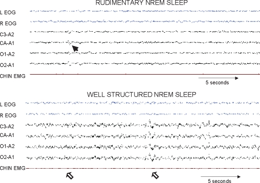
Objectives: In critically ill patients, sleep derangements are reported to be severe using Rechtschaffen and Kales (R&K) methodology; however, whether such methodology can reliably assess sleep during critical illness is unknown. We set out to determine the reproducibility of 4 different sleep-assessment methods (3 manual and 1 computer-based) for ventilator-supported critically ill patients and also to quantify the extent to which the reproducibility of the manual methods for measuring sleep differed between critically ill and ambulatory (control) patients.

Design: Observational methodologic study.

Setting: Academic center.

Patients: Critically ill patients receiving mechanical ventilation and age-matched controls underwent polysomnography.

Interventions: None.

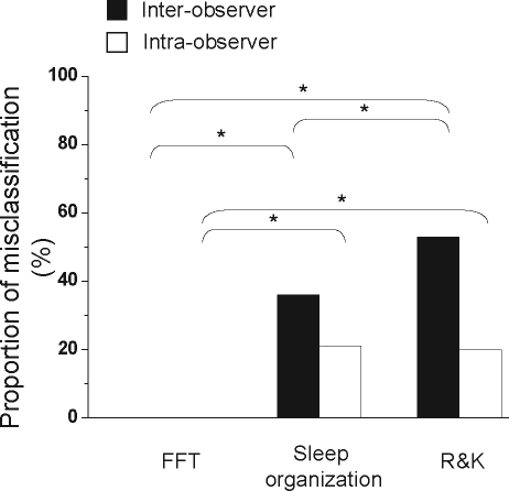
Measurements and results: Reproducibility for the computer-based method (spectral analysis of electroencephalography [EEG]) was better than that for the manual methods: R&K methodology and sleep-wakefulness organization pattern (P = 0.03). In critically ill patients, the proportion of misclassifications for measurements using spectral analysis, sleep-wakefulness organization pattern, and R&K methodology were 0%, 36%, and 53%, respectively (P < 0.0001). The EEG pattern of burst suppression was not observed. Interobserver and intraobserver reliability of the manual sleep-assessment methods for critically ill patients (kappa = 0.52 +/- 0.23) was worse than that for control patients (kappa = 0.89 +/- 0.13; P = 0.03). In critically ill patients, the overall reliability of the R&K methodology was relatively low for assessing sleep (kappa = 0.19), but detection of rapid eye movement sleep revealed good agreement (kappa = 0.70).

Conclusions: Reproducibility for spectral analysis of EEG was better than that for the manual methods: R&K methodology and sleep-wakefulness organization pattern. For assessment of sleep in critically ill patients, the use of spectral analysis, sleep-wakefulness organization state, or rapid eye movement sleep alone may be preferred over the R&K methodology.


Figures

Figure 1
Figure 1
Representative polysomnography tracings of a critically ill patient that show monophasic pattern, characterized by continuous low-voltage theta-delta activity (top panel; 30-second epoch). The bottom panel is a representative tracing from a different critically ill patient that shows cyclic alternating pattern of electroencephalographic activity. Note that this panel is a compressed (60-second) window that reveals low-voltage theta activity alternating (highlighted by horizontal bars) with high-voltage monomorphic delta waves. L EOG refers to left electrooculogram; R EOG, right electrooculogram; C3-A2 and C4-A1, coronal electroencephalograms; O1-A2 and O2-A1, occipital electroencephalograms; EMG, electromyogram.
Figure 2
Figure 2
Representative polysomnography tracings of rudimentary sleep pattern in a critically ill patient as previously described by Valente and colleagues (upper panel). The arrow points to a rudimentary K complex. Representative polysomnographic tracings of a patient with well-formed elements of non-rapid eye movement (NREM) sleep. Note the well-structured K-complex and classic spindles depicted by open arrows (lower panel). Other abbreviations are the same as for Figure 1.
Figure 3
Figure 3
Representative polysomnography tracings of sleep pattern in a critically ill patient that shows rapid-eye movement (REM) sleep (upper and lower panels). Such classic REM sleep alternating with non-REM sleep in critically ill patients were classified as the best sleep organization pattern as previously described by Valente and colleagues. 21 Other abbreviations are the same as for Figure 1.
Figure 4
Figure 4
Interobserver (top panel) and intraobserver (bottom panel) agreement for sleep staging per Rechtschaffen and Kales methodology in critically ill (ICU; open symbols) and ambulatory patients (closed symbols). Columns depict agreements (Cohen κ agreement value) for critically ill (white columns) and control (black columns) patients for all sleep-wakefulness stages by Rechtschaffen and Kales methodology (all), for 4 collapsed groups (light non-rapid eye movement sleep [NREM] stage 1 and 2; slow wave sleep [SWS]; rapid eye movement [REM]; and wakefulness), 3 collapsed groups (NREM, REM, wakefulness). Note that interobserver reliability is worse for critically ill than control patients (top panel; analysis of variance P < 0.0001). Interobserver reliability was worse in critically ill patients than controls for all sleep stages except REM sleep (Neuman-Keuls; *P < 0.05). Also note that the reliability for assessing REM sleep is good in critically ill patients and is not statistically different than that in controls (open symbols; top panel).
Figure 5
Figure 5
Box and whisker plots of the interobserver and intraobserver reliability (Cohen κ agreement value) for assessing sleep in critically ill patients while using spectral analysis by Fast Fourier Transform (FFT), sleep-wakefulness organization pattern (Sleep organization), or Rechtschaffen and Kales (R&K) methodology. The reliability was best for spectral analysis (FFT) when compared with the other methods (Friedman analysis of variance; P = 0.03). Post-hoc (Neuman-Keuls) comparisons revealed a tendency for spectral analysis to be better than sleep-wakefulness organization pattern (P = 0.10) and R&K methodology (P = 0.06).
Figure 6
Figure 6
Proportion of observations (patients or epochs) that were misclassified while assessing sleep in critically ill patients with either spectral analysis by Fast Fourier Transform (FFT), sleep-wakefulness organization pattern (Sleep organization), or Rechtschaffen and Kales (R&K) methodology. Proportion of misclassifications for both interobserver (black columns) and intraobserver (white columns) are shown. For both interobserver and intraobserver reliability testing, the proportion of misclassifications was highest for R&K methodology and least for FFT (2 × 3 χ2 comparisons; P < 0.0001). Significant post-hoc comparisons (2 × 2χ2 comparisons with Bonferroni correction) are also shown (*P < 0.001).

References

    1. Arzt M. Association of sleep-disordered breathing and the occurrence of stroke. Am J Respir Crit Care Med. 2005;172:1447–51. - PMC - PubMed
    1. Gottlieb DJ, et al. Association of usual sleep duration with hypertension: the Sleep Heart Health Study. Sleep. 2006;29:1009–14. - PubMed
    1. Lorenzi-Filho G. Obstructive sleep apnea and atherosclerosis: a new paradigm. Am J Respir Crit Care Med. 2007;175:1219–21. - PubMed
    1. Parthasarathy S. Sleep in the intensive care unit. Intensive Care Med. 2004;30:197–206. - PubMed
    1. Leung RS. Sleep apnea and cardiovascular disease. Am J Respir Crit Care Med. 2001;164:2147–65. - PubMed

MeSH terms