. 2009 Jan;29(1):1-7.
doi: 10.1089/jir.2008.0036.

Evidence for activation of the TGF-beta1 promoter by C/EBPbeta and its modulation by Smads

Affiliations

Evidence for activation of the TGF-beta1 promoter by C/EBPbeta and its modulation by Smads

Selvajothi Abraham et al. J Interferon Cytokine Res. 2009 Jan.

Abstract

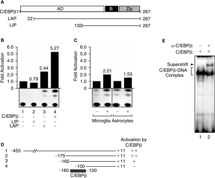
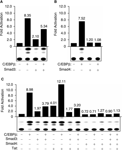
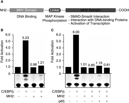
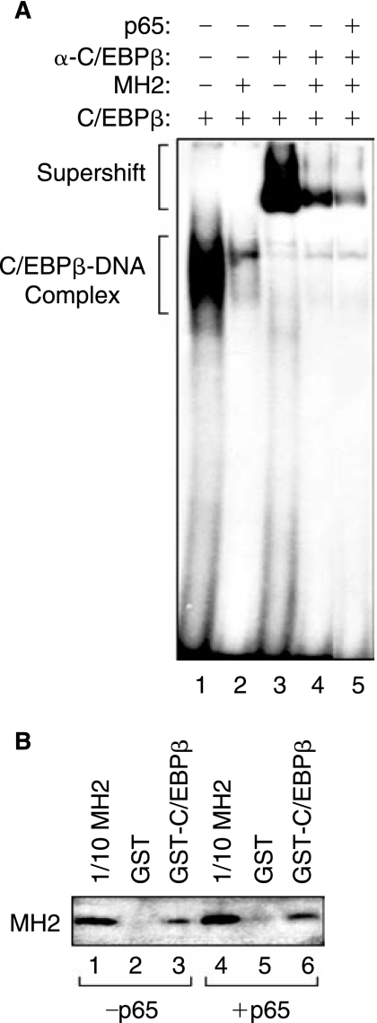
The transforming growth factor-beta1 (TGF-beta1) is a cytokine involved in many biological events inlcuding immunosuppression, angiogenesis, cell growth, and apoptosis. Expression of TGF-beta1 at the transcriptional level is controlled by a series of ubiquitous and specialized factors whose activities can be modulated by a variety of signaling events. Here we demonstrate that activity of the TGF-beta1 promoter is increased by C/EBPbeta, a DNA-binding transcription factor whose activity can be influenced by several immunomodulators, in astrocytes and microglial cells. Interestingly, expression of Smad3 and Smad4, the downstream regulators of the TGF-beta1-signaling pathway, impairs the activity of C/EBPbeta on the TGF-beta1 promoter. Further, we demonstrate that MH2, a common domain among Smads that has protein-binding activities, interacts with C/EBPbeta and decreases its association with a region of the TGF-beta1 promoter that is responsive to C/EBPbeta activation. Interestingly, the p65 subunit of nuclear factor-kappaB (NF-kappaB), which also interacts with C/EBPbeta, cooperates with MH2 and decreased DNA-binding and transcriptional activities of C/EBPbeta on the TGF-beta1 promoter. These observations indicate that an autoregulatory mechanism, involving the MH2 domain of Smads, modulates activation of the TGF-beta1 promoter by C/EBPbeta. Further, our results show that the interplay between NF-kappaB and C/EBPbeta has an impact on the ability of C/EBPbeta to stimulate TGF-beta1 transcription, hence, suggesting that the cross-communication of signaling pathways that modulate NF-kappaB and C/EBPbeta may dictate the level of TGF-beta1 promoter activity.


Figures

FIG. 1.
FIG. 1.
Regulation of TGF-β1 by C/EBPβ. (A) A schematic diagram of C/EBPβ and its deletion mutants LIP and LAP. The human astroglioma cell lines, U-87MG (B and D) or primary human microglia and astrocytes (C), were transfected with 5 μg of TGF-β1-CAT reporter plasmid [full length (B and C) or deletion mutants (D)] alone or cotransfected with 5.0 μg of C/EBPβ, LIP, or LAP expression plasmids, as described previously (Amini and others 2002). The amount of DNA used for transfection was normalized with pcDNA3 plasmid. Cell extracts were prepared 48 h after transfection, and CAT assays were performed, as we have previously described (Sawaya and others 1998). The values shown on the top of each bar represent the fold activation over the basal promoter activity arbitrarily set at one (B and C). The values shown on the right represent significant activation (++), modest activation (+), or no effect (−) of C/EBPβ over the basal promoters (D). The data represent the mean value of at least three separate transfection experiments (SE ± 15%). (E) Gel electrophoretic mobility shift assay (EMSA) was performed, as described previously (Amini and others 2002). Ten micrograms of nuclear extract from C/EBPβ-transfected U-87MG cells (lanes 1 and 2) were used in DNA-binding experiments. For supershift assays, antibodies directed against C/EBPβ (lane 2) (Santa Cruz Biotechnology) were mixed with nuclear proteins for 1 h at 4°C prior to the addition of the probe.
FIG. 2.
FIG. 2.
Functional interplay between C/EBPβ and Smad3/4 in the presence of HIV-1 Tat. U-87MG cells were transfected with 5.0 μg of TGF-β1-CAT reporter plasmid (full length) alone or cotransfected with 5.0 μg of C/EBPβ (A–B), Smad3 (A and C), Smad4 (B and C), or Tat (C) expression plasmids using various combinations. The amount of DNA used for transfection was normalized with pcDNA3 plasmid. Cell extracts were prepared 48 h after transfection, and CAT assays were performed. The values shown on the top of each bar represent the fold activation over the basal promoter activity arbitrarily set at one. The data represent the mean value of at least three separate transfection experiments (SE ± 15%).
FIG. 3.
FIG. 3.
Functional interaction of p65 and MH2 with C/EBPβ. (A) A schematic representation of Smad3 protein and its domains. (B and C) U-87MG cells were transfected with 5 μg of TGF-β1-CAT reporter plasmid alone or cotransfected with 5.0 μg of C/EBPβ, MH2, or p65 expression plasmids using various combinations. The amount of DNA used for transfection was normalized with pcDNA3 plasmid. Cell extracts were prepared 48 h after transfection, and CAT assays were performed. The values shown on the top of each bar represent the fold activation over the basal promoter activity arbitrarily set at one. The data represent the mean value of at least three separate transfection experiments (SE ± 15%).
FIG. 4.
FIG. 4.
Physical association between C/EBPβ and MH2 or p65. (A) Gel electrophoretic mobility shift assay (EMSA) was performed using 10 μg of nuclear extracts from U-87MG cells transfected with C/EBPβ (lanes 1–5) alone, with C/EBPβ + MH2 (lanes 2, 4, and 5), or with C/EBPβ + p65 (lane 5). For supershift assays, antibodies directed against C/EBPβ (lanes 3–5) were mixed with nuclear proteins for 1 h at 4°C prior to the addition of the probe. The positions of the C/EBPβ-DNA complexes are indicated with brackets. (B) GST pull-down. Whole cell extract from cells transfected with 2.5 μg CMV-C/EBPβ was incubated with either glutathione-S-transferase (GST) bound to glutathione beads (lanes 2 and 5) or full length GST-C/EBPβ fusion protein bound to glutathione beads (lanes 3 and 6). Lanes 1 and 4 represent direct analysis of the input protein extract by Western blot, used as a positive control for migration of C/EBPβ on the gel.

References

    1. Abraham S. Sweet T. Sawaya BE. Rappaport J. Khalili K. Amini S. Cooperative interaction of C/EBP beta and Tat modulates MCP-1 gene transcription in astrocytes. J Neuroimmunol. 2005;160:219–227. - PubMed
    1. Amini S. Clavo A. Nadraga Y. Giordano A. Khalili K. Sawaya BE. Interplay between cdk9 and NF-kappaB factors determines the level of HIV-1 gene transcription in astrocytic cells. Oncogene. 2002;21:5797–5803. - PubMed
    1. Coyle-Rink J. Sweet T. Abraham S. Sawaya B. Batuman O. Khalili K. Amini S. Interaction between TGFbeta signaling proteins and C/EBP controls basal and Tat-mediated transcription of HIV-1 LTR in astrocytes. Virology. 2002;299:240–247. - PubMed
    1. Descombes P. Schibler U. A liver-enriched transcriptional activator protein, LAP, and a transcriptional inhibitory protein, LIP, are translated from the same mRNA. Cell. 1991;67:569–579. - PubMed
    1. Eldeen MB. Deshmane SL. Simbiri K. Khalili K. Amini S. Sawaya BE. MH2 domain of Smad3 reduces HIV-1 Tat-induction of cytokine secretion. J Neuroimmunol. 2006;176:174–180. - PubMed

Publication types

MeSH terms

LinkOut - more resources